
«`html
Understanding Innate Immunity and Inflammasomes
The innate immune response constitutes the first line of defense of the immune system, characterized by its immediate and non-specific activation. This robust system includes several essential components for the body's protection:
- Physical barriers, such as the tight junctions of the epithelium and the mucociliary layer that protects the surfaces of the respiratory, gastrointestinal, and genitourinary system.
- Specialized white blood cells that identify and eradicate pathogens without requiring prior specific recognition.
- Membrane-bound and intracellular receptors, such as proteins inflammasomes, which recognize microbial invaders microbes. and trigger a signaling cascade [1].
What is an Inflammasome and What is its Immunological Function?
The inflammasome is an essential protein complex within innate immunity. Its main function is to assemble upon detecting microbes or danger signals, thereby activating a crucial signaling cascade to mount a defensive response.
Several types of inflammasomes have been documented, including [2]:
- The NOD-like receptor (NLR) P3 inflammasome.
- The absent in melanoma 2 (AIM2) inflammasome.
- The NLRC4 inflammasome.
- The pyrin inflammasome.
Structurally, an inflammasome is composed of three main elements [3]:
- A molecular pattern recognition receptor (PRR).
- An adaptor protein, the apoptosis-associated speck-like protein (ASC), which contains a caspase recruitment domain.
- The When acute hives is caused by reactions similar to serum sickness (such as those following blood transfusions or certain medications), it may be accompanied by ecchymosis (bruising), fever, Caspase-1.
Through the secretion of cytokines pro-inflammatory cytokines, notably interleukin (IL)-1β and IL-18, inflammasomes regulate the immune response immunological to both exogenous (e.g.,. bacteria) and endogenous (e.g.,. neoplasia) stimuli [4]. Furthermore, they are fundamental for inducing neutrophil recruitment [4]. neutrophils [4].
Dysregulation of inflammasomes has been associated with significant pathologies, such as acute y chronic inflammatory [4].
What Characterizes an Inflammatory Skin Disease?
Inflammatory skin diseases manifest through the coordinated hyperactivation of the innate and adaptive immune systems, which translates into excessive production of pro-inflammatory mediators, such as cytokines cytokines [4]. These conditions are primarily categorized as defect or syndromes and autoinflammatory.
Cutaneous autoimmune pathologies, exemplified by lupus erythematosus and vitiligo, involve aberrant responses directed against autoantigens. autoantigens. This erroneous response is strongly orchestrated by B cells and B-cell and B and
```
the presence of autoantibodies.
Skin autoinflammatory diseases, such as periodic fever syndromes and neutrophilic dermatoses, fever and dermatosis neutrophilic, involve the activation of innate immune system cells (macrophages, neutrophils, mast cells and natural killer [NK] cells), which causes tissue damage in the absence of autoantigens and autoantibodies.
Furthermore, certain skin conditions, such as psoriasis, present both autoimmune and autoinflammatory components. [4].
We will use the examples of dermatitis allergic contact dermatitis and psoriasis to illustrate the fundamental role that inflammasomes play in chronic inflammatory skin diseases.
Inflammasome-Mediated Skin Conditions
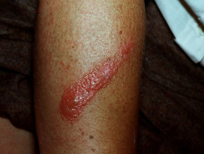
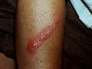
Allergic Contact Dermatitis
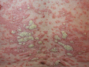
Chronic Plaque Psoriasis
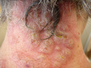
Acute neutrophilic dermatosis
The Role of Inflammasomes in Allergic Contact Dermatitis
Allergic contact dermatitis manifests as a rash pruritic y lesions..., itchy, and erythematous rash, resulting from a renal hypersensitivity reaction to a small molecule [5]. molecule [5].
In experimental models of contact dermatitis induced by 2,4-dinitrofluorobenzene (DNFB), it has been observed that the activation of the adaptive immune system is intrinsically linked to the prior activation of the innate immune system. [5].
The justification for the production of IL-1 family cytokines mediated by inflammasomes in contact dermatitis is based on the following evidence:
- The essential components of the inflammasome are present within the keratinocytes. [6].
- Exposure to DNFB leads to the secretion of IL-1β in these animal models. [6].
- Antagonists of the IL1 receptor (IL-1RA) have demonstrated therapeutic remarkable effects in the treatment of contact dermatitis. [4].
Inflammasomes and the Pathophysiology of Psoriasis
Psoriasis is a prevalent skin pathology defined by the appearance of well-demarcated erythematous plaques, covered by a silvery scale. plaques erythematous well-demarcated plaques, covered by a The presence of a superficial silvery scale.
The pathophysiology The pathophysiology.
- The immune cells. Innate immune cells orchestrate the activation of T helper (Th)-1 and Th-17 lymphocytes, which are responsible for releasing key pro-inflammatory cytokines such as IL-1β and IL-18. [8,9].
- The elevated expression of IL-1β and IL-18 is upregulated via caspase-1 and the AIM-2 pathway within keratinocytes. [8,10].
- The sustained presence of these inflammatory cytokines causes intensified recruitment of immune cells, abnormal keratinocyte proliferation, and proliferation abnormal of keratinocytes and a inflammation chronic inflammation. [8].
Currently, detailed research is being conducted on their precise implication in this disease, seeking new therapeutic targets.
antagonists of IL-1 family members for the treatment of psoriasis [2].
What Defines an Autoinflammatory Disease?
Autoinflammatory syndromes and diseases manifest through recurrent inflammatory episodes, triggered by a hyperactive innate immune system response. The [1,3]. key pathogenesis in these conditions does not involve the presence of specific autoantibodies or the participation of T cells.
To diagnose an autoinflammatory disease, it is essential to first rule out underlying infectious, neoplastic or allergic conditions.
Among the recognized autoinflammatory syndromes are hereditary periodic fevers, hereditary,, other monogenic autoinflammatory syndromes defined, as well as acquired or polygenic non-hereditary disorders (exemplifying, generalized pustular psoriasis) nail psoriasis pustular generalized) and associated neutrophilic dermatoses.
The Role of the Inflammasome in Autoinflammatory Dermatosis
Protein complexes known as inflammasomes positively regulate the signaling of various cytokines, including interleukin-1 (IL-1), in the context of autoinflammatory skin diseases. This upregulation amplifies the neutrophil-mediated inflammatory cascade through various mechanisms. Specifically, IL-1β exerts anti-apoptotic effects on neutrophils, resulting in a prolongation of their lifespan.
- Inflammasome assembly is initiated when Pattern Recognition Receptors (PRRs) detect stimuli and promote interaction with the adaptor molecule ASC, thus achieving caspase-1 activation [5].
- Caspase-1, once activated, processes and releases the cytokines IL-1β and IL-18, which are potent stimulators of the inflammatory response [5].
- These pro-inflammatory cytokines activate neutrophils, which subsequently will infiltrate infiltrate the skin and other organs. This infiltration causes specific skin symptoms such as pustules, ulceration, pustules, ulceration or rashes, in addition to manifesting systemic symptoms steroids such as low-grade fever and general malaise. malaise allergy.
Understanding inflammasome activation is crucial for developing targeted therapies, such as the aforementioned IL-1 antagonists, which seek to interrupt this chronic inflammatory cycle in dermatological conditions.


