
Key Histopathological Findings in Herpes Virus Infections
The histological manifestations observed in infections caused by herpes simplex virus, varicella, and herpes zoster present notable similarities. Establishing a definitive differential diagnosis requires meticulously correlating these clinical findings with specialized laboratory analyses, such as immunohistochemistry, viral culture, or PCR testing, using an appropriate tissue sample or swab.
Distinctive Histopathological Features of Herpes Diseases
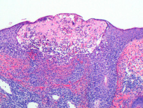
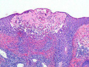
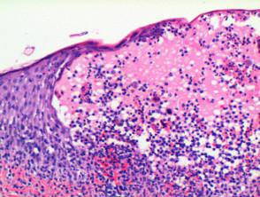
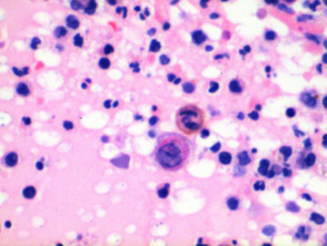
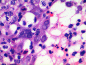
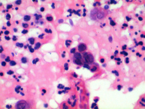
The characteristic histopathological presentation of herpetic infections allows for a reliable diagnosis. When examining the sample at low power, a typical lesion reveals the formation of an intraepidermal blister (see Figure 1). The fundamental diagnostic finding is dyskeratotic, acantholysis keratinocytes , evidenced by the presence of individual keratinocytes floating freely within the blister cavity (Figures 1, 2, 3).
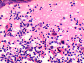
Affected keratinocytes exhibit clear nuclear alterations, resulting from viral replication. These changes include the margination of the chromatin chromatin multinucleated and the appearance of intranuclear toward the nuclear periphery, the development of multinucleated nuclei, and the appearance of intranuclear inclusions (Figures 3, 4, 5). These viral inclusions typically appear as basophilic/pink deposits surrounded by a clear halo within the nucleus; nucleus; when observed along with the other nuclear modifications typical of herpes, they are classified as Cowdry Type A inclusions. It should be noted that Cowdry Type B inclusions are associated with other viruses, such as poliovirus, and do not possess these specific nuclear characteristics of herpesviridae.
In the initial phases of the pathological process, cytoplasmic vacuolization changes may be observed, especially notable in the basal layer cytoplasm, of the epidermal cells. As these cells swell and separate, the cytoplasm acquires a predominantly eosinophilic stain, which is especially marked in the cell multinucleated cells. The inflammatory infiltrate eosinophilic, eosinophils multinucleated cells.. The infiltrate inflammatory that accompanies the lesion is mixed in nature, dominated by lymphocytes and neutrophils, with a common presence of lymphocytes y neutrophils, scattered eosinophils (Figure 6).
Detailed Pathology of Herpes Virus Infection
To fully understand the spectrum of these viral lesions, it is vital to continue exploring the images and their specific histopathological correlates.
Histopathological Analysis of Herpetic Lesions
Herpetic folliculitis can manifest with very subtle histological changes, making it easily underestimated, particularly in the early stages or when the follicle is only partially involved. The key finding is a **necrotic hair follicle** surrounded by a dense superficial and deep lymphocytic infiltration, along with the presence of neutrophils. The characteristic cytopathic signs of the virus are distinguished in the acantholytic cells of the epithelium, located predominantly in the infundibular region or in the adjacent affected epidermis.
In cases of varicella or verrucous herpes zoster infection, in addition to the expected viral cytopathic alterations, the concurrence of epidermal hyperkeratosis and acanthosis is observed.
Advanced Diagnostic Methods for Herpes Infections
To accurately differentiate between herpes simplex virus type 1 (HSV-1), type 2 (HSV-2), and varicella zoster virus (VZV), immunoperoxidase staining using a specific monoclonal antibody is recommended. Likewise, to confirm the precise etiology of the infection, it is feasible to process cultures or perform Polymerase Chain Reaction (PCR) on the pertinent clinical sample, which allows for definitive distinction between HSV and VZV.
Differential Diagnosis Criteria
The differential diagnosis of herpetic lesions is crucial, as it can be confused with other skin pathologies presenting with vesicles or ulcers, such as:
- Erosions associated with pyoderma or secondary impetigo.
- Erythema multiforme.
- Severe cutaneous candidiasis.
- Drug reactions.
A correct correlation between clinical findings (such as lesion distribution), detailed histopathological presentation, and the results of molecular or culture tests will ensure an accurate diagnosis and appropriate antiviral treatment for each variant of herpetic infection.
Differential Diagnosis of Herpes Infections
The differential diagnosis of vesicular lesions caused by herpes simplex virus (HSV) requires the exclusion of other skin pathologies that present with vesicles and blisters. Several conditions that must be considered are detailed below:
Differentiation from Viral Infections and Autoimmune Pathologies
Coxsackie Virus Infection / Hand, Foot, and Mouth Disease: The vesicles associated with this condition, caused by enteroviruses such as Coxsackie, show vesiculation intraepidermal vesiculation and signs of acantholysis, but crucially, they lack the characteristic nuclear inclusions or the formation of multinucleated giant cells seen in true herpetic infections.
Pemphigus Vulgaris: Although Pemphigus Vulgaris (PV) can generate blisters, its clinical and pathological presentation differs significantly. In PV, suprabasal acantholysis is observed, and in late lesions, blister formation occurs. In the acantholytic space, few cells detach, and most importantly, the viral cytopathic changes are not appreciated. However, it is essential to recognize a serious complication: a dissemination of HSV (Kaposi's chickenpox-like eruption) can overlap with a pre-existing case of pemphigus vulgaris or vice versa. In these complex diagnostic scenarios, direct At the dermal-epidermal junction, an increase in the density of dermal papillae is observed, which are surrounded by a bright, monomorphic layer of cells., suprabasal acantholysis vesicles. vesicles. immunofluorescence immunofluorescence often proves diagnostic. If viral etiology needs confirmation, the specialized molecular tests mentioned previously should be applied.
Post-Herpetic Sequelae and Associated Pathologies
It is pertinent to mention that the site where a herpes zoster (HZ) episode previously occurred has been associated with the subsequent appearance of various conditions. These include autoimmune and neoplastic conditions such as chronic lymphocytic leukemia, granuloma annulare, lichen planus, leukemia lymphocytic chronic, Sweet's granuloma keloid scars, Avoidance Strategies for Triggering Factors, vasculitis, lymphoma lichen planus reactions typical manifestations of active viral infection are rarely observed in the presentation of these late sequelae.
To achieve an accurate diagnosis in cases of herpetic infections, differentiating these secondary presentations and mimicking pathologies is essential to determine the most appropriate antiviral or immunosuppressive treatment.


