
Fibrosarcoma was diagnosed much more frequently before the implementation of immunohistochemical studies. Currently, it is estimated that this neoplasm accounts for less than 1% of all sarcomas adult sarcomas. These tumors have the capacity to arise de novo (de novo) or develop within another type of sarcoma sarcoma already present.
Histology and Presentation of Fibrosarcoma
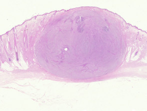
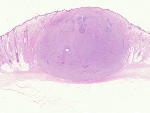
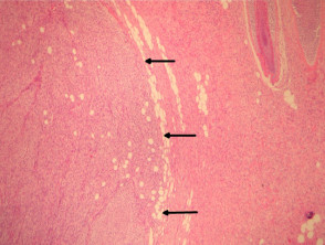
In fibrosarcoma, histological sections reveal a dermal (Figure 1) or subcutaneously cellular infiltrate composed of spindle cells. subcutaneous tumor composed of spindle cells. It is important to note that the tumor can appear de novo or in association with another sarcoma. Figure 2 illustrates a fibrosarcoma (left side) arising within a dermatofibrosarcoma protuberans (right side), with arrows delineating the invasion border.
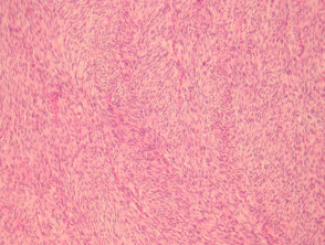
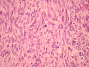
The tumor component is made up of spindle cells exhibiting relative uniformity and a marked affinity for basophilic dyes basophils (Figures 2 to 4). Characteristically, the malignant cells and even are organized following a pattern reminiscent of a «herringbone» (the pattern is more evident in Figure 3). Mitotic figures (in the tumor area, this finding is not accompanied by evidence of significant cytological) are typically easily observable (Figure 4).
There are two well-defined morphological variants: the infantile variant and the epithelioid epithelioid sclerosing variant. Fortunately, the occurrence of these variants in the skin is extremely rare.
Fibrosarcoma Pathology Images
Key Special Studies for Fibrosarcoma Diagnosis
It is essential to use an exhaustive immunohistochemical panel to rule out other types of tumors malignancies. Fibrosarcoma is generally negative for S100 (which excludes melanoma), also shows negativity for keratins (with exceptions such as melanomacarcinoma and synovial sarcoma), carcinoma and smooth muscle markers (which rules out leiomyosarcoma) synovial biopsy) leiomyosarcomaand CD34 (which helps differentiate it from dermatofibrosarcoma protuberans).
Essential Differential Diagnosis of Fibrosarcoma
As detailed previously, the rigorous application of immunohistochemical studies is indispensable for establishing clear distinctions from other sarcomas that present similar morphologies. Performing this immunological profiling is the most critical step to arrive at the mucous membranes. correct diagnosis of fibrosarcoma.


