
Deciphering Erythropoietic Protoporphyria: Characteristics and Histological Findings
Erythropoietic protoporphyria (EPP) is a metabolic disorder caused by a deficiency of the ferrochelatase **enzyme**, resulting in an **intracellular** accumulation of heme group precursors. Clinically, it manifests with **acute** **photosensitivity** starting in childhood, causing **erythema** and **scaling**, which eventually leads to scarring and a characteristic waxy thickening of the skin.
Histopathology of Erythropoietic Protoporphyria
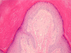
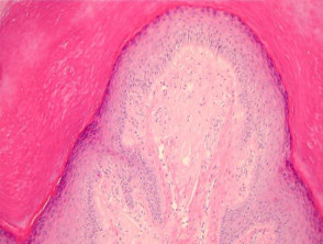
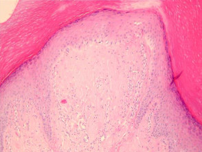
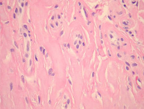
Under the microscope, a deposit of **hyaline** **eosinophilic** material is observed in the **dermis**, specifically around the walls of the **blood vessels**. This involvement can be extensive, encompassing the surrounding dermis and mimicking colloid **milia** (see figures 1 and 2). Increased magnification reveals a notable thickening of the vascular structures (figure 3).
In early EPP lesions, similar to porphyria cutanea tarda, **subepidermal** bullae may appear. These bullae show scant cellularity and a **scant inflammatory** **infiltrate**. In the **basal** layer of the **epidermis** overlying these bullae, it is possible to identify «caterpillar bodies,» which are segmented **linear** structures composed of degenerated **keratinocytes** and **basement membrane** components.
Pathological Correlation in Erythropoietic Protoporphyria
Specialized Studies in Erythropoietic Protoporphyria
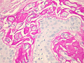
The hyaline material surrounding the **blood vessels** reacts positively to PAS staining (figure 4) and shows resistance to diastase. Direct immunofluorescence demonstrates the deposition of **immunoglobulin** (mainly IgG), **fibrin**, and C3 around the blood vessels in the **papillary dermis**. Additionally, the presence of the membrane attack complex (C5-9) has been detected in the vascular walls. This material also stains favorably with Sudan black and Hale's colloidal iron methods, confirming the nature of the deposits.
Accurate understanding of these histopathological features is crucial for the differential diagnosis and effective management of erythropoietic protoporphyria, especially when evaluating the extent of skin damage and therapeutic response.
Caterpillar bodies are PAS positive.
Differential Diagnosis of Erythropoietic Protoporphyria
Porphyria cutanea tarda: Histological similarities are notable. Typically, the vascular deposits vascular in porphyria cutanea tarda are less evident than in EPP and are limited to the vessel wall. Solar elastosis elastosis (frequent in PCT due to patient age) does not characterize EPP.
SclerodermaScleroderma: The dermal alterations dermal of chronic EPP chronic can mimic scleroderma; however, the collagen collagen in EPP exhibits a looser texture. Bullae and vascular cuffs are not distinctive features of scleroderma.
Lipoide Proteinosis: The vascular cuff is present in both pathologies; however, in EPP, this cuff does not extend to the adnexal structures Reflectance Confocal Microscopy (RCM) allows for the classification of skin lesions into main categories, facilitating their differential diagnosis. These categories include:. Clinical correlation is extremely useful for distinction.
Colloid Milia: EPP shows great similarity to colloid milia when the accumulation in the papillary dermis is extensive (figures 1, 2). In colloid milia, fissures form fissures within the material and vascular thickening is less pronounced.
The differentiation Differentiation from other conditions with scant-cell subepidermal bullae (such as epidermolysis bullosa, pseudoporphyria, and bullous amyloidosis , scabies, bullous disease) is established through clinical features, immunofluorescence analysis, and tests for porphyrins in serum porphyrin gel sickness, feces, and urine.
Distinguishing Erythropoietic Protoporphyria from other dermatoses requires an exhaustive analysis of clinical and laboratory findings. Diagnostic confirmation is strongly supported by precise measurements of body porphyrin levels.


