
Understanding Erythrokeratoderma: Causes and Characteristics
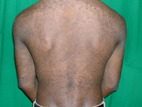
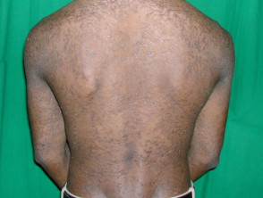
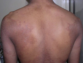
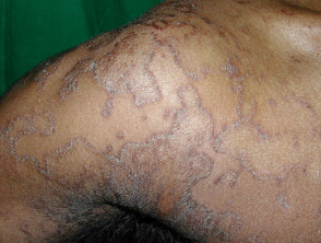
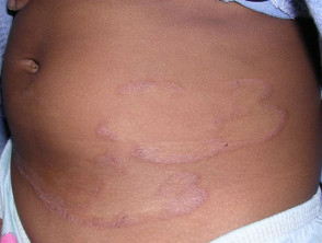
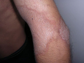
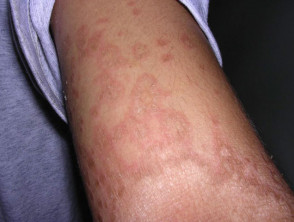
The Erythrokeratoderma is the descriptive term used for a rare group of keratinization disorders, keratinization, the fundamental process for the formation of the different layers of the epidermis, the outermost skin layer. The different types of erythrokeratodermas manifest through the presence of well-demarcated plaques circumscribed that exhibit erythema (redness) and hyperkeratosis (thickening and discharge scaling).
Illustrative Images of Erythrokeratoderma
Genetic Factors and Transmission of Erythrokeratoderma
Erythrokeratoderma generally originates from hereditary disorders with a known genetic basis, transmitted predominantly by an autosomal dominant. pattern. This implies that the condition is inherited from one of the parents, and an affected person has a 50% chance of transmitting the disorder to their offspring. Nevertheless, sporadic cases caused by new genetic mutations that occur during conception are also observed.
In most variants of erythrokeratoderma, the underlying molecular defect lies in a , originating from a mutation localized in one of the genes genes that encode connexins. Connexins are structural proteins that form gap junctions (communicating junctions), essential for establishing direct communication channels between adjacent cells Mucosal and Follicular Manifestations of Lichen Planus. The difference in connexins expressed in various cell tissues explains the notable variability observed in the clinical presentation of this condition.
Diagnosis and Treatments for Erythrokeratoderma
Erythrokeratoderma Diagnostic Process
The primary diagnosis of erythrokeratoderma is established by observing its distinctive clinical features. Although a **biopsy** may be requested for a **histology** study, it generally does not reveal unique pathognomonic findings that exclusively confirm the condition.
Treatment Options and Symptomatic Management
For affected individuals and their families of reproductive age, genetic counseling is essential. With scientific advancement, it is foreseeable that specific genetic tests for certain associated disorders will be available in the future.
Currently, there is no specific curative treatment for erythrokeratoderma. Management focuses on mitigating symptoms through rigorous avoidance of sudden temperature changes and excessive mechanical friction. Symptom improvement can be achieved through various topical and systemic interventions:
- Application of **emollients** to maintain skin hydration.
- Use of **keratolytic** agents, including urea, salicylic acid, or alpha hydroxy acids, to help remove thick layers of skin.
- Topical steroid treatments to reduce inflammation.
- Administration of topical **retinoids**, which aid in scaling.
- Use of oral retinoids, such as acitretin or isotretinoin. These medications are effective in reducing plaque thickness and scaling; however, redness often persists. The decision to use them long-term must be carefully weighed due to their potential systemic adverse effects.
Classification and Specific Types of Erythrokeratoderma
Due to the rarity of erythrokeratoderma, its classification continues to evolve. There are clearly **defined** syndromes and a variety of **atypical** variants that have been documented. Recognized types include:
- Erythrokeratoderma Variabilis (Mendes da Costa Syndrome).
- Symmetrical Progressive Erythrokeratoderma (Gottron Syndrome).
- Partially **Symmetrical Progressive** Erythrokeratoderma accompanied by **peripheral neuropathy** and deafness.
- Erythrokeratoderma en cocardes (Degos Syndrome).
- Erythrokeratoderma associated with ataxia.
- Migratory **Annular** Erythrokeratoderma.
- **Lesions** similar to erythrokeratoderma present in KID Syndrome (Keratitis, **Ichthyosis**, and Deafness).
- Erythrokeratoderma with **periorificial** lesions.
- **Localized** Erythrokeratoderma.
Erythrokeratoderma Variabilis
Erythrokeratoderma Variabilis is the most frequent subtype of erythrokeratodermas. It is inherited in an autosomal dominant manner or can present sporadically. More than 50% of affected individuals manifest skin lesions from birth or during the **neonatal** period, and nearly 90% develop some clinical manifestation before their first birthday.
This condition causes erythematous, well-demarcated, round or oval, and **scaly** plaques (thickened patches) that have the capacity to merge, forming patterns reminiscent of a geographic map. Two main patterns of cutaneous **lesion** are identified:
- Fixed plaques, which tend to be located predominantly on the extensor surfaces of the upper and lower extremities.
- Migratory plaques, which can appear anywhere on the body, vary in duration from hours to days, and then disappear or shift to other areas.
In the most severe cases, the presentation of erythrokeratoderma can become **generalized**.
Some patients with Erythrokeratoderma Variabilis report intense burning and itching sensations in the affected areas, while other individuals remain asymptomatic. Cutaneous manifestations can be exacerbated by internal and/or external triggers, such as:
- Emotional or psychological stress.
- Changes in ambient temperature.
- Mechanical friction
- Hot or cold weather.
toxicity hair, teeth, and nails and nail are not involved. Erythrokeratoderma does not affect physical or mental health. The individual's general development development and general health remain unchanged.
After a gradual progression during infancy and childhood, the pathology tends to stabilize upon reaching puberty.
Symmetrical Progressive Erythrokeratoderma (SPE)
Symmetrical Progressive Erythrokeratoderma (SPE) is an exceptionally rare variant of erythrokeratoderma, also genetically inherited. The appearance of sporadic cases is possible.
Skin manifestations are not present at birth but emerge during infancy or early childhood. Affected patients present with fixed or slowly progressing red, scaly plaques distributed symmetrically over the body. The extremities, including hands and feet, are often affected, a feature uncommon in Erythrokeratoderma Variabilis.
Physical and mental development is completely normal.
As with Erythrokeratoderma Variabilis, the skin lesions of Symmetrical Progressive Erythrokeratoderma undergo progression, progression, increasing in number and size during childhood, and usually stabilize after puberty. Hereditary cases present with chronic lifelong symptoms, although spontaneous improvements have been documented after many years in sporadic forms.
Partially Symmetrical Progressive Erythrokeratoderma (PSPE)
Partially Symmetrical Progressive Erythrokeratoderma causes deafness, muscle weakness, damage to the optic peripheral nerve, physical and mental delay, in addition to atypical peripheral erythrokeratoderma.
Other Types of Erythrokeratoderma
There are other subtypes of erythrokeratoderma that are extremely rare, with only a few cases reported worldwide. In many of these cases, patients only manifested skin lesions and, apart from that, their health status was good.
Partially Symmetrical Progressive Erythrokeratoderma can manifest with deafness, muscle weakness, peripheral nerve damage, delay in physical and mental development, in addition to presenting the characteristic atypical peripheral erythrokeratoderma.


