
Erythema Nodosum: General Description and Histological Findings
The Erythema Nodosum is considered the most frequent manifestation of inflammation of the subcutaneous fat tissue).. It typically presents as with elevated, painful and tender to the touch, subcutaneous nodules nodules y plaques subcutaneous, which emerge with greater predilection in the pretibial areas pretibial grooves bilaterally.
Key Histopathology of Erythema Nodosum
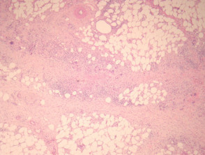
From a histological perspective histological, Erythema Nodosum represents the archetype of a septal panniculitis. The characteristic histopathological presentation is that of a septal panniculitis with a mixed inflammatory component. This includes a infiltrate cellular cellular infiltrate composed of lymphocytes, and a smaller proportion of histiocytes., giant cells, lymphocytes, histiocytes, giant cells, and occasionally eosinophils eosinophils, notably highlighting the absence of vasculitis Avoidance Strategies for Triggering Factors (see Figures 1 to 3).
It is postulated that Miescher's radial granulomas—small nodules formed by spindle-shaped or oval histiocytes arranged around tiny clefts—constitute a fairly specific finding in Erythema Nodosum (see Figure 4).
When the lesions When lesions first appear, it can be complex to make a diagnosis based solely on histopathological criteria. Initial sections may reveal edematous septa harboring a inflammatory prominent inflammatory infiltrate and minimal presence of fibrosis. fibrosis. As the pathology progresses, a neutrophilic infiltration is observed, followed by chronic infiltrates, the development of granulomas infiltration infiltration, neutrophilic infiltration telangiectasias Risk of blindness in extreme cases, granulomas granulomas , and finally, septal fibrosis.
It is important to mention that small-vessel vasculitis (or venulitis) can be observed in the initial stages of the disease.
Pathological Morphology of Erythema Nodosum
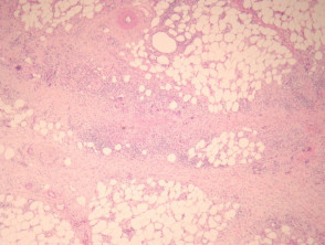
Figure 1
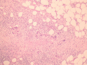
Figure 2
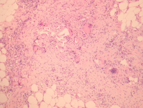
Figure 4
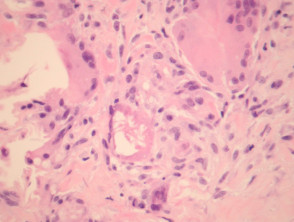
Figure 5
Additional Investigations for Erythema Nodosum
Generally, no additional diagnostic procedures are required. However, if the neutrophilic infiltrates are very intense, staining could be considered to identify possible causative microorganisms. microorganisms Implementing rigorous sun protection measures from an early age to significantly reduce the risk of skin damage and the development of skin cancers.


