
Erythema Elevatum Diutinum: Characteristics and Histopathological Findings
The Erythema elevatum diutinum (EED) is a dermatological condition chronic and rare. It typically manifests through the appearance of papules y nodules reddish-brown papules and nodules. These lesions are usually located on the extensor surfaces, especially on the back dorsal of the hands. In addition to skin manifestations, it is common for patients to present with systemic symptoms steroids, strains, such as arthralgia. Its association with various hematologic y rheumatologic, which underscores the need for comprehensive evaluation.
Detailed Histopathology of Erythema Elevatum Diutinum
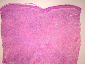
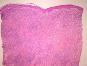
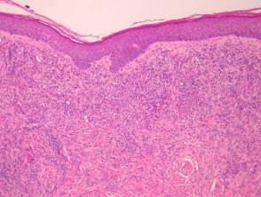
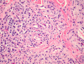
The histology of erythema elevatum diutinum reveals characteristic findings in the dermis. It is important to note the persistence (preservation) of the papillary dermis and the and (seen in Figures 1 and 2). Microscopically, a infiltrate vascular is observed that affects the upper and middle dermis. This infiltrate is predominantly composed of neutrophils, although lymphocytes, eosinophils y plasma cells are also identified in smaller proportions (Figures 3, 4, 5).
In the most recent lesions, it is common to identify leukocytoclasia (Figure 5). As the lesions evolve, dermal systemic sclerosis occurs, accompanied by the persistent presence of plasma cells and residual foci of Avoidance Strategies for Triggering Factors leukocytoclastic vasculitis. In some cases, this sclerosis process can be marked and manifest with a storiform pattern (Figures 1 and 4), representing a distinctive morphological characteristic of this dermatosis. dermatosis.
Images Correlating the Pathology of Erythema Elevatum Diutinum
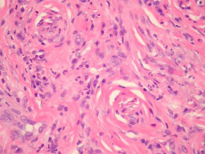
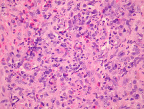
Understanding the clinical presentation of erythema elevatum diutinum, along with its distinctive findings on skin biopsy, is crucial for the accurate diagnosis of this rare vasculitis. The correlation between cutaneous signs and possible systemic comorbidities should always be considered in patient management.
Detailed Differential Diagnosis of Erythema Elevatum Diutinum
To establish an accurate diagnosis of Erythema Elevatum Diutinum (EED), it is crucial to distinguish it from other skin conditions with similar clinical presentations. The key conditions for differential diagnosis are detailed below:
Conditions to Differentiate from Erythema Elevatum Diutinum
Granuloma Faciale: Some experts consider this condition to be a manifestation of the same disorder, but limited to the facial region. Both clinical processes share characteristics of small-vessel sclerosing vasculitis. It is important to note that granuloma faciale generally presents a lower association with systemic disease compared to EED.
Leukocytoclastic Vasculitis: The most acute forms of leukocytoclastic vasculitis exhibit a less dense infiltrate cell infiltrate. Clinically, these forms tend to show greater red blood cell extravasation extravasation compared to established EED.
Acute Neutrophilic Dermatoses: Although clear clinical distinctions exist, the initial stages of EED lesions can resemble Sweet's Syndrome and other dermatosis characterized by neutrophilic predominance.
Dermatofibroma: The later and fibrotic fibrotic phases of Erythema Elevatum Diutinum can mimic a dermatofibroma. However, EED lesions lack the characteristic pattern of peripheral collagen collagen trapping and hyperplasia Other conditions that cause significant edema in the superficial dermis include mild epidermal hyperplasia typically seen in dermatofibroma.
Eosinophilic Angiocentric Fibrosis: This condition is distinguished by the presence of fibrosis (which can occasionally adopt a storiform pattern) surrounding small arteries, vessels, commonly located in the orbit and upper respiratory tract. It is accompanied by telangiectasias and inflammatory mixed inflammatory infiltrates with a marked predominance of eosinophils. Recently, a possible association between this entity and elevated IgG4 levels has been documented.
For a definitive diagnosis, the correlation between clinical presentation and histopathological findings is fundamental to differentiate Erythema Elevatum Diutinum from these similar pathologies, ensuring the most appropriate treatment for the patient.


