Understanding Erosive Pustular Dermatosis
What is Erosive Pustular Dermatosis?
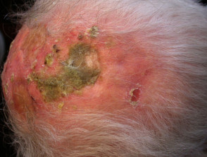
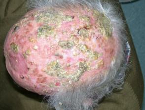
Erosive pustular dermatosis manifests through localized areas characterized by pustules, lakes of pus, or crusts, overlying eroded plaques or nodules. Predominantly affecting sun-damaged scalp in elderly individuals, this condition presents a distinctive clinical picture.
This disorder is also known by the terms erosive pustulosis or erosive pustular dermatosis of the scalp, making it crucial to differentiate it from other skin pathologies.
Demographic Profile and Risk Factors
Erosive pustular dermatosis affects both men and women. Most diagnosed individuals are over 80 years old and frequently present with actinic keratosis, along with a history of squamous cell carcinoma or, less commonly, basal cell carcinoma on the scalp. Typically, the condition emerges in areas of abnormal healing, such as those resulting from skin cancer surgery or previous herpes zoster episodes.
As another neutrophilic dermatosis, it may be observed with greater incidence in patients with immunosuppression, rheumatoid arthritis, and myeloid hematological disorders.
Additionally, erosive pustular dermatosis has been documented as an adverse side effect associated with the use of epidermal growth factor receptor inhibitors, such as gefitinib.
Etiology of Erosive Pustular Dermatosis of the Scalp
The precise cause of erosive pustular dermatosis of the scalp remains unknown. However, a strong correlation with sun damage exposure is established.
Frequently, the disorder is triggered by minor skin trauma in the affected area (including surgical procedures), a phenomenon known as pathergy, and may manifest with poor wound healing. Infection is not considered the primary etiological factor, given that lesions do not resolve solely with the administration of antibiotics.
When the condition is located on the lower extremities, circulatory problems such as venous stasis and associated edema may be involved.
Key Clinical Manifestations
Erosive pustular dermatosis typically begins with minute pustules appearing on the scalp, forehead, or temples. Although less common, localizations on the lower legs have also been reported.
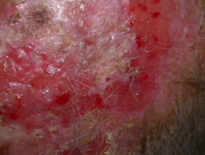
These pustules evolve into the formation of lakes of pus or crusts that present shades varying between brownish-yellow and greenish. If these crusts detach, the underlying dermis is revealed as red and exudative. When the disease is extensive, it can progress to permanent scarring and significant alopecia.
Erosive Pustular Dermatosis: Complications and Diagnosis

Erosive Pustular Dermatosis
Erosive Pustular Dermatosis
Erosive Pustular Dermatosis
Complications Associated with Erosive Pustular Dermatosis
Erosive Pustular Dermatosis, being a chronic inflammatory condition, carries risks of significant complications. The main problems that may arise include:
- Skin alterations associated with this condition may include: hair (Hair loss (alopecia)), which can be transient or result in permanent baldness.
- Secondary bacterial infection bacterial of the eroded areas.
- Elevated risk of developing skin cancer over chronically affected areas.
Diagnostic Methods for Erosive Pustular Dermatosis
The primary diagnosis of erosive pustular dermatosis is established through clinical observation of its distinctive characteristics. However, complementary tests are required to rule out other etiologies and confirm suspicion.
The Bacterial cultures obtained via swabs can identify the presence of Staphylococcus aureus. Likewise, it is prudent to perform mycological scrapings to exclude fungal infections such as tinea capitis or kerion.
Due to the clinical difficulty in differentiating the underlying erosion from a possible skin carcinoma, a biopsy skin biopsy of the eroded skin is essential. Performing multiple and deep biopsies biopsies is usually very useful to obtain a definitive diagnosis. Histopathology analysis Histopathology typically reveals subcorneal and non-follicular pustules subungual and non infiltrate, thickening at the level of the. Furthermore, epidermal hypertrophy or atrophy along with erosions can be observed. hypertrophy o limb epidermal hypertrophy or atrophy along with and erosions. erosions. In the dermis, a inflammatory mixed inflammatory infiltrate is found, comprising plasma cells y neutrophils, plasma cells and neutrophils, although these histopathological findings frequently lack specificity.
of thrombophilic lymphocytic arteritis? mucous membranes. What is the Differential Diagnosis of Erosive Pustular Dermatosis?
When evaluating Erosive Pustular Dermatosis, it is crucial to consider a broad spectrum of skin pathologies that may present similar clinical features. Conditions to keep in mind for the differential diagnosis include:
- Merkel cell squamous cells, Squamous cell carcinoma, or other types of skin cancer.
- Details of the Laboratory Procedure mucosa Mucous membrane pemphigoid, Brunsting-Perry type.
- Infection.
A correct and thorough diagnostic process is essential to ensure that treatment is directed appropriately to this complex dermatological condition, thereby minimizing long-term complications.
- Bacterial or fungal infection.
- Folliculitis decalvans.
Treatment of Erosive Pustular Dermatosis
To treat the lesions, crusts should be removed with a gentle wash, possibly using potassium permanganate (Condy's crystals) or a solution of acetic acid (diluted vinegar) as an antiseptic astringent. Subsequently, a soft dressing is applied over the wound.
Any secondary bacterial infection should be addressed with antistaphylococcal oral antibiotics, such as flucloxacillin or erythromycin.
Erosive pustular dermatosis of the scalp responds well to the application of potent or ultrapotent steroids once or twice daily on the affected areas for a couple of weeks. If the skin disorder recurs, treatment should be repeated as necessary. If long-term topical steroid use is required, a calcineurin inhibitor, such as tacrolimus ointment, might be more appropriate.
Other treatments that have proven useful include:
- Anti-inflammatory antibiotics for six weeks or more (e.g., minocycline).
- Calcipotriol cream to reduce scaling.
- Cryotherapy for underlying actinic keratoses.
- Topical dapsone.
- Zinc sulfate.
- Photodynamic therapy.
- Photodynamic therapy.
Oral retinoids, such as acitretin and isotretinoin.
Prevention of Erosive Pustular Dermatosis.
Patients diagnosed with erosive pustular dermatosis must rigorously protect themselves from the sun (using wide-brimmed hats when outdoors) and be vigilant in recognizing early signs of recurrence, which should be treated immediately according to the established regimen.
New actinic keratoses and cutaneous carcinomas, especially squamous cell carcinoma, can develop in the affected areas. It is crucial to diagnose and treat them without delay. Since they can be difficult to differentiate from erosive pustular dermatosis of the scalp, repeated biopsies may be necessary to confirm the diagnosis.
Prognosis and Outlook for Erosive Pustular Dermatosis.