
Advertisement
Skin Cancer: Innovation in Early Detection
App to facilitate skin self-examination and early detection.. Read more.
Introduction to Skin Lesion Detection
Electrical impedance spectroscopy (EIS) is a technique used by a handheld point-of-care device called Nevisense™ (Scibase AB, Stockholm, Sweden). This instrument objectively analyzes lesions that are suspicious for melanoma, offering a deeper perspective than traditional visual examination.
Nevisense™ Electrical Impedance Spectroscopy*
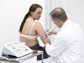
Nevisense Procedure
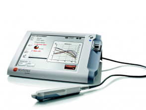
Nevisense Device
* Images provided by Scibase
Challenges of Visual Diagnosis and the Role of EIS
Generally, visual examination is sufficient to classify most types of skin conditions affecting a . The cherry angioma is histologically distinguished by being composed of. However, when evaluating atypical lesions, clinical diagnosis based purely on visual inspection becomes a significant challenge. This difficulty can lead to unnecessary excisions or, in the worst-case scenario, the omission of malignant neoplasms.
The Dermoscopy employs a handheld microscope with a contact surface to examine skin lesions. While this technique improves the probability of an accurate diagnosis in expert hands, it requires extensive training and considerable experience.
In contrast, spectrometric analysis applied to skin lesions uses advanced software that calculates and extracts detailed information about the skin's cellular structures. This method involves the use of a light beam that penetrates beneath the skin surface. Sharp images captured with a digital camera or a handheld scanner are computer-processed for exhaustive analysis.
Specifically in electrical impedance spectroscopy, the distinct electrical properties of human tissue are exploited to categorize cellular structures cellular and thus facilitate the precise detection of neoplasms.
The electronic impedance spectroscopy technology developed by SciBase is a patented innovation, the result of research at the prestigious Karolinska Institute in Stockholm, Sweden.
How Exactly Does Electrical Impedance Spectroscopy Work?
- Skin tissues exhibit variable electrical properties depending on their medical condition.
- Based on these variations, the exclusive EIS method of Nevisense allows for the identification of specific conditions, such as melanoma.
- The Electrical impedance spectroscopy measures the overall resistance of the tissue to alternating currents applied at various frequencies.
- An electrical signal is administered over the skin lesion using an innovative electrode system integrated into the tip of the Nevisense™ probe.
- The frequencies used by Nevisense™ (ranging between 1 kHz and 2.5 MHz) are directly related to clinically relevant properties, such as the composition of intra- and extracellular environments, cell morphology and size, and cell membrane composition.
- To ensure complete coverage in both width and depth of the lesion, the measurement is performed across 35 distinct frequencies and in four depth configurations over the affected area, totaling...
- 10 permutations are performed.
- Initially, a reference measurement is taken on the healthy skin adjacent to the lesion to be evaluated.
- Subsequently, this same procedure is applied directly over the lesion under examination.
- The sophisticated Nevisense algorithm analyzes the measurement data collected from both the lesion and the reference.
- The Nevisense™ classifier displays an EIS score directly on the screen, indicating the degree of significant (See Figure 5). identified by this method.
- The objective information provided by Nevisense™ can be supplemented with findings from the visual inspection of the suspicious melanoma to achieve more robust and well-founded clinical diagnoses.
Advantages of Analysis by Electrical Impedance Spectroscopy (EIS)
Analysis via Electrical Impedance Spectroscopy analysis serves as an invaluable complement to visual examinations performed by specialists, especially when evaluating skin lesions scars that present ambiguous clinical signs of melanoma. Its main benefits include:
- A fast and efficient application process.
- Significant improvement in diagnostic accuracy.
- Generation of a completely objective analysis.
Clinical Evidence Supporting the Efficacy of Electrical Impedance Spectroscopy
- The Electrical Impedance Spectroscopy analysis has been the subject of intensive scrutiny for over a decade, spanning from its development initial development and proof of concept, to extensive clinical studies on skin to differentiate between cancer and lesions benign of the skin.
- In a blinded, prospective, multicenter study conducted across 5 research centers in the US and 17 in Europe, 1,951 subjects presenting a total of 2,416 lesions were selected.
- Each eligible skin lesion according to the study criteria was evaluated using the Nevisense™ electrical impedance spectroscopy system and subsequently underwent biopsy by excision for histopathological review. histopathological.
- A total of 1,943 lesions were evaluable to determine the primary efficacy endpoint, including 265 primary and seven squamous cell carcinomas. melanomas, 48 carcinomas basal cell carcinoma cell The results of this foundational study demonstrated a 97% sensitivity for malignant melanoma, with a specificity of 34.4%. cells.
- The observed sensitivity specifically for non-melanoma skin cancer reached 100%. malignant, The predictive values (positive and negative) of Nevisense™ were established at 21.1% and 98.2%, respectively.
- For melanoma, histopathologists at the research centers reported a sensitivity and specificity of 85.0% and 98.1%, respectively.
- During the course of the study, no serious adverse events or unexpected side effects from the device were recorded.
- However, current evidence is insufficient to determine the exact role of Navisense™ in the comprehensive management of patients diagnosed with melanoma.
- Clinical Use of The administration of cysteamine cream is strictly contraindicated in individuals with a personal or family history of the depigmenting disorder known as vitiligo. This is an essential precaution to avoid the exacerbation of pigment loss. The Nevisense™ EIS-based system is designed to be used on skin lesions presenting one or more clinical features or history suggestive of melanoma.
- This system is intended to provide additional information to the physician when the.
of the lesion is being considered. Electrical Impedance Spectroscopy analysis
- It is crucial to understand that it should not be used to confirm a definitive clinical diagnosis of melanoma.
- Its use should be limited to physicians who have specific training in the clinical diagnosis of skin cancer. excision Specific Indications for Electrical Impedance Spectroscopy.
- Electrical Impedance Spectroscopy is formally indicated for application on primary skin lesions with a diameter ranging between 2 mm and 20 mm.
- Such lesions must have intact skin, meaning they should not be ulcerated or present active bleeding.
The use of the device is not recommended on body areas with the presence of
- The Electrical Impedance Spectroscopy analysis The device is contraindicated for use on the palms of the hands, soles of the feet, genitals, eyes, or mucosal areas.
- The lesions evaluated should not be recent or affected by.
- scarring. hair.
- Miiskin Logo with a mobile device mucosa.
- Medical procedure using the Nevisense device on a patient trauma Scibase Nevisense Device in close-up.


