
Eccrine Hidrocystoma: Definition and Dermatological Histology
The **eccrine hidrocystoma** is classified as a . The cherry angioma is histologically distinguished by being composed of cystic, whose most accepted genesis lies in the cystic retention of the nodes eccrine. Nevertheless, there is debate among some medical authorities suggesting a possible apocrine origin for this clinical entity.
Detailed Histology of Eccrine Hidrocystoma
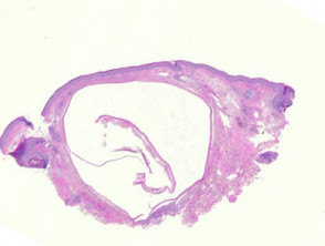
Histopathologically, a cystic space occupying the dermis, is observed, which may present as unilocular (see Figure 1). This cystic space is lined by a thin epithelial layer, . Occasionally, transepidermal elimination and marked, typically composed of one or two cell layers. The cells exhibit a cytoplasm slightly eosinophilic cytoplasm and a bilaminar structure (illustrated in Figures 2).
A distinctive characteristic in this lesion is the absence of prominent luminal dilations, commonly referred to as ‘apocrine snouts’.
Pathology and Microscopic Characteristics of Eccrine Hidrocystoma
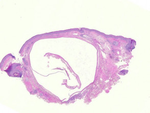
Figure 1
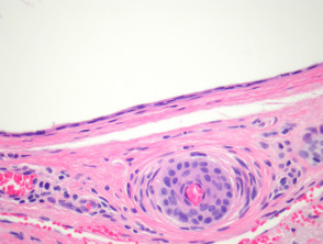
Figure 2
Complementary Examinations for Eccrine Hidrocystoma
Diagnostic studies are generally not required to confirm eccrine hidrocystoma, given its characteristic clinical presentation and histopathological findings.
Differential Diagnosis in the Pathology of Eccrine Hidrocystoma
It is essential to distinguish eccrine hidrocystoma from apocrine hidrocystoma. Although certain interpretations suggest an apocrine origin for the eccrine type, the epithelium epithelium of apocrine hidrocystoma often exhibits notable luminal dilations, i.e., the aforementioned 'apocrine snouts', which is absent in the eccrine variant, thus facilitating the diagnosis..


