
Key Histopathological Features of Darier Disease
Darier disease is classified within the group of acantholytic dyskeratotic lesions. Its hallmark is the presence of suprabasal separation, At the dermal-epidermal junction, an increase in the density of dermal papillae is observed, which are surrounded by a bright, monomorphic layer of cells., resulting from a process of dyskeratotic accompanied by focal dyskeratosis of the keratinocyte.
Histological Analysis of Darier Disease
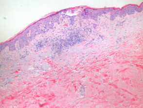
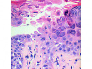
The histological study of Darier disease reveals an epidermal process epidermal and superficially dermal of an inflammatory inflammatory (Figure 1). At this magnification, intraepidermal separation can be observed, intraepidermal, which upon closer examination proves to be suprabasal acantholysis (Figures 2 and 3). This acantholysis is detectable at all levels of the epidermis.
Keratinocyte dyskeratosis is evidenced by two notable morphological alterations. Corps ronds (Corps ronds) are characterized by the presence of small nuclei. pyknotic nuclei, a clear perinuclear halo perinuclear and an cytoplasm eosinophilic (Figures 4 and 5).
On the other hand, grains, which are flattened cells with elongated nuclei, are prominently located in the stratum corneum stratum corneum and the granular layer granulomatous (Figures 4 and 5).
The epidermis in the upper layers shows orthokeratosis compact, which can occasionally manifest as a prominent focal plug.
In the dermis superficial dermis, the most common finding is a perivascular lymphocytic infiltrate lymphocytic perivascular of mild intensity (Figure 2). The presence of eosinophils is infrequent. eosinophils is infrequent.
Visualization of the Pathology of Darier Disease
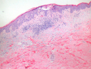
Figure 1
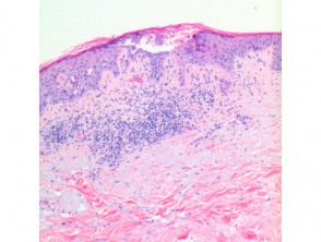
Figure 2
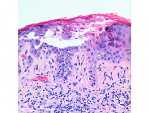
figure 3
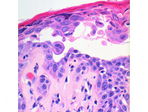
Figure 5
Special Stains and Syndromes Associated with Sebaceoma
Precise understanding of these histopathological features is fundamental to confirm the clinical diagnosis of Darier disease and differentiate it from other keratinocytic pathologies. The combination of suprabasal acantholysis and focal dyskeratosis defines the characteristic cellular picture of this skin condition.
Histological Variants of Darier Disease
In the comedonal variant of Darier disease, the areas of acantholytic dyskeratosis are more pronounced, presenting prominent keratotic plugs and extensive hyperplastic formations keratotic prominent and extensive formations that are hyperplastic. of the epidermis that extend deep into the superficial dermis.
In bullous Darier disease, the distinction lies mainly in the size of the acantholytic fissures fissures acantholytic, which create lacunae containing scattered acantholytic cells within them.
Hopf's acrokeratosis verruciformis, although its classification as a component of Darier disease or a separate entity remains debated, is included due to its clinical association. The lesions lesions show papillomatosis church steeple-shaped papillomatosis, along with hyperkeratosis y acanthosis epidermal hyperkeratosis and acanthosis. Upon examining very thin sections, it is possible to identify small foci of acantholytic dyskeratosis.
Differential Diagnosis of Darier Disease
To differentiate it from Hailey-Hailey disease, the key is the extent of acantholysis affecting the epidermis. In Hailey-Hailey disease, more extensive areas of cellular involvement, both partial and total, are observed.
In the case of dermatosis Transient transient transient acantholytic dermatosis (Grover's disease), although the Darier-type variant can replicate almost identical characteristics, the presence of smaller involved foci and an inflammatory infiltrate that includes eosinophils helps guide the diagnosis toward Grover's disease.
Accurate histopathological diagnosis is essential to differentiate these conditions and ensure the most appropriate dermatological treatment for each patient.


