
«`html
Cutaneous Manifestations of Hematologic Diseases
Hematologic diseases encompass a vast spectrum of disorders affecting the chemical constituents fundamental components of the blood. This includes cellular conditions—red blood cells (erythrocytes), white blood cells (leukocytes), and platelet—as well as malignant neoplasms originating from these cell lines. Identifying the cutaneous signs associated with these hematologic conditions is crucial, as they are often key to early diagnosis or represent direct complications of the underlying pathology.
Anemia: Dermatologic Signs of Cellular Deficiencies
Nutritional Anemia and Its Signs on the Skin
Deficiency of essential nutrients such as iron, vitamin B12, or folate constitutes a primary cause of anemia, manifesting clearly in the skin and mucous membranes.
The characteristic cutaneous sequelae of these deficiencies include:
- Pallor marked in the conjunctiva (mucous membranes) and in the palm creases. palm of the hand.
- nails., which presents as a smooth, reddened-looking tongue.
- Development of poikilodermatous hypopigmentation.
- In cases of B12 and folate deficiency, hyperpigmentation, serving as a possible sign differential diagnosis.
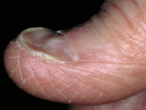
- Increased nail fragility lesion, frequently observed as koilonychia (spoon-shaped nail deformity).
Exploring Key Cutaneous Signs in Anemia
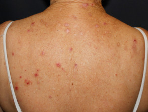
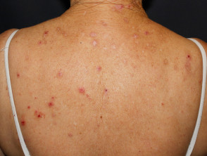
Pruritus associated with iron deficiency
Presence of sparse eyelashes.
Hemolytic Anemia and Its Complications
Hemolytic anemia, exemplified by sickle cell anemia, arises from accelerated destruction of red blood cells, triggering a series of secondary clinical manifestations:
- Experience of Pruritus intense skin pruritus.
- Development of Jaundice, resulting from increased bilirubin levels.
- Possible appearance of Petechiae and hemosiderin deposits, visible as hemosiderosis (small macules brown macules on the skin).
Myeloproliferative Disorders and Dermatologic Manifestations
Hematopoiesis, the fundamental process of blood cell formation, gives rise to two main cell lineages: lymphoid or myeloid. Myeloproliferative neoplasms (MPNs) specifically affect this latter pathway, generating abnormal proliferation of bone marrow cells. lymphoid or the myeloid. Myeloproliferative neoplasms (MPNs) specifically affect this latter pathway, generating an abnormal proliferation of bone marrow cells.
Polycythemia Vera: A Chronic Myeloproliferative Disorder
Polycythemia vera is a prototypical example of a chronic chronic myeloproliferative disorder. This condition is characterized by the uncontrolled overproduction of blood components by the myeloid bone marrow, frequently causing a marked increase in the red blood cell count.
The dermatologic manifestations associated with Polycythemia Vera are distinctive and include:
- Plethora or rubicund cyanosis: an excessively flushed complexion due to circulatory overload.
- Aquagenic Pruritus: an intense itching sensation triggered after skin contact with water.
- Erythromelalgia: burning pain and intense redness, predominantly in the hands and feet.
- Livedo Reticularis: a violaceous skin discoloration adopting a reticular or lace-like pattern.
- Acrocyanosis: persistent bluish-violet discoloration in the distal extremities, such as hands and feet.
- Pyoderma gangrenosum: in severe cases, serious skin ulcerations may appear.
Images of Cutaneous Manifestations in Polycythemia Vera
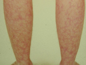
Livedo Reticularis
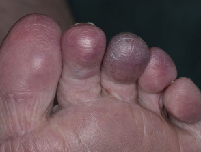
Acral Cyanosis
Host-versus- Mast Cell (Mastocytosis)
Mast cell disease, or mastocytosis, arises from a myeloid disorder characterized by the pathological accumulation of mast cells mast cells in various organs and tissues of the body. This condition is primarily classified into two types: cutaneous mastocytosis, more prevalent in the pediatric population, and systemic mastocytosis systemic sclerosis), which predominates in adults.
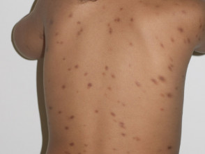
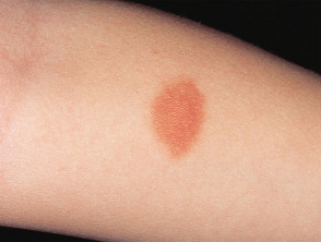
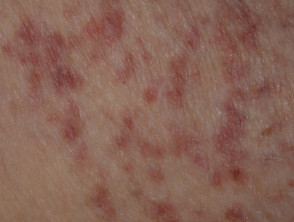
The cutaneous manifestations associated with mastocytosis are highly suggestive and typically present in the following ways:
- Maculopapular cutaneous maculopapular: It manifests as papules of a tan or brown color in children, and as reddish-brown macules and papules in adults. Clinically, this form is known as hives Pigmentary incontinence.
- Darier's Sign: Immediate skin reaction characterized by the appearance of erythema and wheals (hives) after friction or rubbing of the affected skin.
- Generalized pruritus: Diffuse and often severe itching.
- Mastocytomas: Nodular lesions that are observed less frequently, especially in adult patients.
Visual Examples of Cutaneous Signs of Mastocytosis
Leukemia Cutis: Dermatologic Presentation
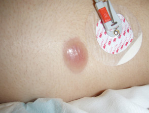
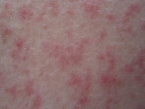
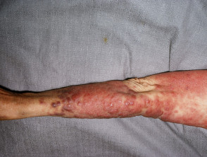
Leukemia cutis is a rare dermatologic presentation that emerges in the context of myeloproliferative disorders that remain undiagnosed or untreated. This includes pathologies such as acute myeloid leukemia acute (AML) and other Lymphoproliferative.
Upon detection of these cutaneous manifestations associated with myeloproliferative diseases, it is essential to seek specialized medical evaluation. This ensures diagnostic confirmation and the initiation of appropriate treatment, given the possible progression of the underlying conditions.
When infiltration infiltration of the skin by leukemic cells occurs, it is termed lymphoma skin. This infiltration manifests through papules, nodules y plaques with a highly variable morphology. Commonly, these lesions exhibit a coloration ranging from pink, violaceous, purplish or tones more pigmented than the surrounding healthy skin. A relevant aspect is that these manifestations are usually painless to palpation and have a notably hardened.
Illustrative Images of Leukemia Cutis
Cutaneous Lymphoproliferative Disorders
Lymphoproliferative disorders originate from alterations in the lymphoid lineage of hematopoietic stem cells. Both systemic forms of lymphoma, including Hodgkin's Lymphoma and Non-Hodgkin's Lymphoma, have the capacity to manifest through specific dermatologic signs and symptoms. Below are the cutaneous conditions derived from these lymphomatous processes.
Cutaneous T-Cell Lymphoma (CTCL)
Cutaneous T-Cell Lymphoma (CTCL) represents a type of lymphoproliferative disorder whose primary and predominant location is the skin. The most frequent variant within this group of malignancies is mycosis fungoides. Its distinctive clinical manifestations include:
- Mycosis fungoides: This form is characterized by the appearance of macules y patch test with elevated (reddish coloration) and papillomas., patches, which may present as isolated or multiple lesions. The color spectrum of these lesions can vary notably, ranging from orange to intense red or violet.
- Sézary Syndrome: It manifests as erythroderma o dermatitis Exfoliative ulceration.
These cutaneous conditions are often accompanied by intense secondary symptoms, including debilitating pruritus, diffuse alopecia (hair loss), development of keratoderma palmoplantar —a marked thickening of the skin on palms and soles—and predisposition to Unlike other bacterial superimposed infections.
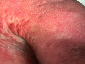
Cutaneous Lymphomas: Classification and Presentation
Sézary Syndrome
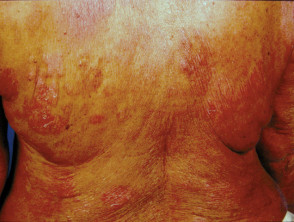
Mycosis fungoides
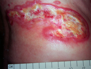
Cutaneous B-Cell Lymphoma (PCBCL)
Primary cutaneous B-cell lymphomas (PCBCLs) represent a group of lymphoproliferative disorders affecting the skin without evidence of dissemination to other organs at the time of diagnosis. Among these is primary cutaneous marginal zone lymphoma.
It is important to note that some subtypes of PCBCL have been etiologically linked to the Epstein-Barr virus (EBV). A particularly aggressive variant requiring high diagnostic suspicion is primary cutaneous diffuse large B-cell lymphoma, leg type.
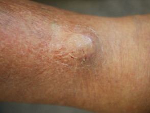
In cases of the leg subtype, patients are usually elderly and present with solitary or grouped plaques or tumors. These lesions are often ulcerated, present a bluish or erythematous coloration, and are mainly located on the lower extremities. Due to their morphology, this presentation can be clinically confused with chronic ulcers secondary to venous insufficiency.
Visual Correlation: Manifestations of Cutaneous B-Cell Lymphoma
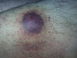
Cutaneous B-cell lymphoma
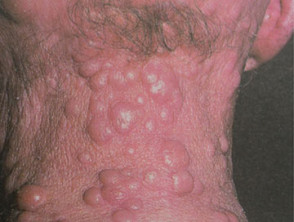
Cutaneous B-cell lymphoma
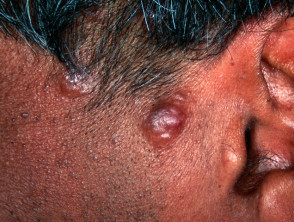
Cutaneous B-cell lymphoma
Plasma Cell Disorders and Cutaneous Immunoglobulinopathies
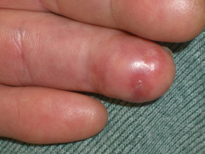
Multiple myeloma, although primarily a hematologic neoplasm, can manifest cutaneously through the rare presentation of cutaneous plasmacytoma.
A relevant systemic syndrome is POEMS syndrome, which involves a complex of five features: polyneuropathy, organomegaly, endocrinopathy, M protein, and skin changes. In this entity, plasma cell proliferation leads to the overproduction of cytokines and other inflammatory mediators that drive the clinical signs.
Additionally, may also occur. Internal organ involvement becomes more likely if the vasculitis is associated with are immunoglobulins that have the capacity to precipitate in the cold. This precipitation is the underlying cause of palpable purpura and hyperviscosity syndrome, frequently associated with Raynaud's Phenomenon. Cryoglobulinemia is classified by its etiology as follows:
- Type 1: Directly associated with underlying B-cell malignancy.
- Mixed (Combining Type 1 with Type II): Related to the presence of infectious agents or autoimmunity.
Proper identification of these manifestations and their correct classification are crucial for establishing the prognosis and the most effective therapeutic strategy in lymphomas and plasma cell dyscrasias.
- B-cell malignancy or autoimmune diseases.
The cold agglutinins are cold-sensitive immunoglobulins that react against antigens present on red blood cells. These reactions can trigger circulatory conditions such as Raynaud's phenomenon and acrocyanosis.
Cutaneous Manifestations of Plasma Cell Diseases

Amyloidosis: Spectrum and Cutaneous Expressions
Amyloidosis comprises a series of disorders defined by the misfolding of proteins, leading to their pathological accumulation in different tissues and organs, including the dermis. The most common subtype is AL amyloidosis, related to monoclonal light chains.
The dermatologic manifestations associated with this condition are clinically relevant and include:
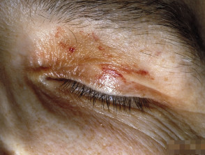
- Petechiae: Small, pinpoint hemorrhages that do not blanch upon pressure.
- Pinch purpura: Notorious hemorrhagic lesions that develop after applying friction or a light pinch to the skin.
- Ecchymosis: Flat bruises of greater extent, with a bluish or violaceous coloration.
- Skin hardening with a waxy texture.
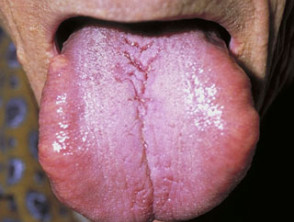
- Macroglossia: Visible and abnormal enlargement of the tongue.
Distinctive Cutaneous Signs in the Diagnosis of Amyloidosis
Hematopoietic Stem Cell Transplantation (HSCT)
Stem cells for transplantation can be collected from peripheral blood, bone marrow, or umbilical cord blood. umbilical. This procedure is considered an essential therapeutic line for aggressive hematologic neoplasms when other options have failed, including diffuse large B-cell lymphoma and certain myeloproliferative disorders.
Complications associated with HSCT arise both from the side effects of the required immunosuppression and from the host's immune response against donor cells, known as Graft-versus-Host Disease (GVHD). Although GVHD, both acute and chronic, can affect virtually any organ (including systems like the liver, lung, and joints), the skin is the site most frequently affected.
The dermal manifestations typically observed after transplantation include:
- Focal erythema over palms, soles, and earlobes.
- Development of bullae or the formation of blisters.
- Folliculitis eosinophilic.
- Poikiloderma, which is characterized by skin thinning (atrophy) combined with uneven pigmentation (hyperpigmentation).
Understanding these cutaneous manifestations is fundamental, as they often represent the first signs of serious post-transplant systemic complications.
- Loss of pigment).
The management and understanding of these dermatologic sequelae are crucial for optimizing long-term outcomes in patients undergoing stem cell transplants. Continuous monitoring by the clinical team is essential to identify and treat any adverse cutaneous reaction early, including chronic chronic GVHD.
Extracorporeal photopheresis (ECP) is used in the treatment of cutaneous GVHD in patients who have received a transplantation stem cell transplant.
Coagulation Disorders and Their Cutaneous Manifestations
Coagulation disorders are defined by an abnormal tendency that can manifest as either excessive clot formation (thrombosis) or uncontrolled bleeding.
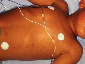
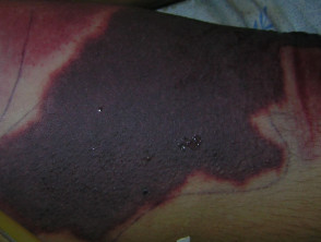
Disseminated Intravascular Coagulation Progressive Disseminated Intravascular (DIC) promotes uncontrolled thrombus formation. This syndrome can be triggered by severe infections, such as meningococcal disease, or underlying pathologies like advanced cancer cancer. In its most severe forms, DIC depletes all available coagulation factors, paradoxically triggering severe hemorrhage.
The dermatologic manifestations associated with DIC include:
- Petechiae.
- Ecchymoses.
- Active bleeding or oozing at sites of wounds, IV access, or catheters.
The Thrombocytopenia, defined as a decrease in platelet count, significantly increases the tendency to bleed. For example, in immune thrombocytopenia, platelet levels platelet are reduced due to the action of undetectable circulating antibodies directed against them. Other causes of thrombocytopenia include bone marrow diseases, Wiskott-Aldrich syndrome, and the administration of certain medications.
The specific cutaneous manifestations of thrombocytopenia include:
- Oral or To rule out other.
- Extensive ecchymoses.
- Purpura with a characteristic pigmented.
There are other coagulation disorders that also present clinically relevant cutaneous signs:
- Antiphospholipid syndrome.
- Necrosis Warfarin-induced skin necrosis.
- Blue toe syndrome.
Blue toe syndrome.
Meningococcal Disease
Disseminated Intravascular Coagulation
The correct identification of these cutaneous signs in infectious and hematologic disorders is vital for the timely management of coagulopathies. Dermatologic surveillance offers early clues regarding the patient's systemic severity.
Cutaneous Manifestations in Inflammatory Disorders Associated with Hematologic Disease
Inflammatory conditions linked to hematologic diseases can manifest on the skin in various ways, signaling possible underlying systemic complications. These manifestations include:
- Hyperpigmentation generalized.
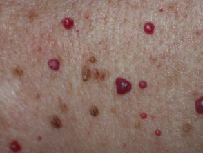
- Proliferative (a tumor originating in the blood vessels).
- Hypertrichosis (excessive hair growth).
- Telangiectasia (visible dilation of small capillaries with an arachnoid or violaceous appearance).
- Vasculitis Hematological and serological analyses commonly reveal the following patterns:.
Sweet's syndrome is also considered one of these cutaneous manifestations linked to hematologic processes. It is crucial that the diagnosis and management of these lesions are approached in a coordinated manner between dermatologists and hematology specialists to ensure comprehensive and effective treatment for the patient.
Acute Neutrophilic Dermatosis and Cutaneous Conditions Associated with Hematology
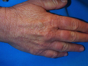
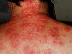
The Acute febrile infiltration neutrophilic dermatosis (known as Sweet's syndrome) presents as an inflammatory skin eruption of sudden onset, clinically characterized by the presence of fever and an increase in the white blood cell count (or leukocytosis). This syndrome can be associated with multiple primary conditions, such as infections, Unlike other, inflammatory bowel disease, certain hematologic neoplasms, and the ingestion of certain pharmacological agents, such as certain antibiotics. A localized presentation of this disease is called dorsal hand neutrophilic dermatosis. dorsal back of the hands.
Dermatologic Characteristics of Sweet's Syndrome
Clinically, Sweet's syndrome exhibits a number of very specific dermatologic characteristics:
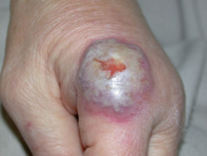
- Emergence of firm papules or nodules, varying in color from bright red to violaceous; they can present as isolated or multiple lesions. In some cases, bullous lesions may appear, or they may manifest similarly to ulcerated pyoderma gangrenosum or clinical mimics of this. bullous, or manifest similarly to ulcerated pyoderma gangrenosum or clinical mimics of this.
- Observable presence of pustules (pus-filled tissue or).
- Appearance of tender erythematous nodules, located predominantly on the extremities, which are sometimes indistinguishable from erythema nodosum.
- Demonstration of the pathergy phenomenon, where dermatologic lesions arise from sites of minimal trauma trauma.
Inflammatory Skin Disease Associated with Hematologic Condition
Adverse Drug Reactions in Hematologic Treatments
Quite frequently, pharmacological agents used to manage certain hematologic conditions can cause toxicities whose main expression is at the cutaneous level. Among the most recurrent examples are:
- Interferon alpha, a treatment used in the management of chronic myeloid leukemia (CML), has the capacity to induce adverse cutaneous manifestations.
- Cutaneous cutaneous porphyria and vitiligo.
- toxicity and of kinase inhibitor, Tyrosine kinase inhibitors, such as imatinib (used in chronic myeloid leukemia - CML and cutaneous mastocytosis), in addition to treatment for sclerodermoid graft-versus-host disease (GVHD), can induce psoriasiform dermatitis, It is common to observe prominent blood vessels directly surrounding these malignant cell groupings., intense itching (pruritus), alopecia and increased sensitivity to sunlight (reactions.).
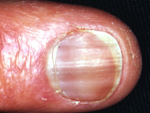
- The use of hydroxyurea frequently causes significant skin dryness, which can trigger the appearance of actinic keratosis, the development of squamous cell carcinoma, chronic lower extremity ulcers, generalized hyperpigmentation, and various nail alterations. seborrheic keratosis actinic, granulomas carcinoma scaly, actinic keratosis and nail.
Cutaneous Adverse Reactions Associated with Hematologic Drugs
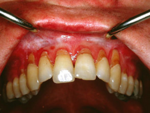
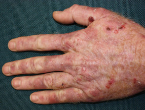
Understanding these potential dermatologic manifestations linked to therapies in hematology is essential for offering comprehensive and effective management to the patient. This understanding allows for a clear distinction between medication-induced toxicities and other primary cutaneous pathologies. Therefore, rigorous dermatologic surveillance is a fundamental practice during the administration of highly complex systemic treatments.


