
Colloid Milium: Clinical and Histopathological Characteristics
The colloid milium is an infrequent dermatosis manifested by the appearance of cupuliform and translucent papules in the dermis. This disorder is linked to the accumulation of dermal colloidal material. There are four recognized variants of this condition: adult colloid milium, juvenile, pigmented, and nodular.
Histological Analysis of Colloid Milium
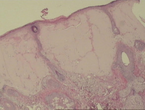
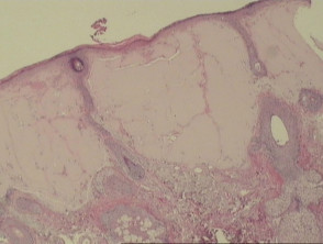
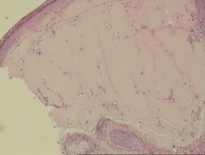
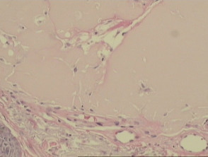
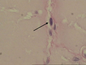
Under histopathological examination, masses of homogeneous, eosinophilic colloidal material are observed, situated in the papillary dermis (visible in Figures 1, 2, and 3). These colloid masses frequently present clefts or fissures, which, on occasion, may be delimited by fibroblasts (indicated by the arrow in Figure 4).
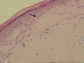
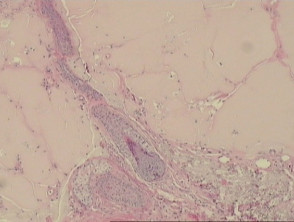
Specifically in adult variant colloid milium, it is common to identify a subepidermal Grenz band that respects the papillary dermis (arrow in Figure 5), along with evidence of solar elastosis. The scarcity of adnexal structures in this form of the disease is notable (Figure 6).
Pathological Visualization of Colloid Milium
Specific Histopathological Analysis for Colloid Milium
In histopathological study, staining with PAS (Periodic Acid of Schiff) is positive for this condition. Additionally, Congo Red and Crystal Violet stains may also show positive results.
Key Differential Diagnosis of Colloid Milium
Primary Cutaneous Amyloidosis: Distinguishing this entity can be complex and often requires the use of specialized histochemical stains. Unlike amyloidosis, colloid milium tests negative for laminin and type IV collagen staining. Cytokeratin is positive in lichenoid amyloidosis, which can aid in differentiation, as colloid milium stains negatively. However, nodular amyloid and cutaneous involvement by systemic amyloid are also negative for cytokeratin. It is important to note that juvenile colloid milium may stain positively for cytokeratin, and some experts consider it a variation of lichenoid amyloidosis. Electron microscopy distinguishes the entities: amyloidosis reveals straight, unbranched filaments, whereas colloid milium filaments are shorter, branched, and wavy.
Erythropoietic Protoporphyria: In these cases, hyaline deposits are observed around and within the walls of blood vessels. Involvement of the dermoepidermal junction may be detected, a feature absent in colloid milium.
Lipoid Proteinosis: Dermal deposits are notably concentrated around blood vessels. Additional distinctive features include atrophy of eccrine sweat glands and involvement of the pilosebaceous units. Unlike colloid milium, in this condition, staining is positive for laminin, type IV collagen, and cytokeratin.
Corneal Conjunctivitis (Woody): Juvenile colloid milium has been documented to be related to this chronic form of pseudomembranous conjunctivitis. A possible common etiologic cause could be plasminogen deficiency. The histopathology of woody conjunctivitis shows a thinned or eroded conjunctival epithelium, amorphous subepithelial eosinophilic masses, and foci of granulation tissue.


