

Understanding Basal Cell Carcinoma (BCC)
The Basal Cell Carcinoma (BCC), known by its technical designation as Basal Cell Carcinoma without melanoma, represents the most frequent form of skin cancer. This cancer is characterized by being a tumor locally destructive tumor, presenting a wide variety of clinical manifestations and histologic.
Detailed Histology of Basal Cell Carcinoma
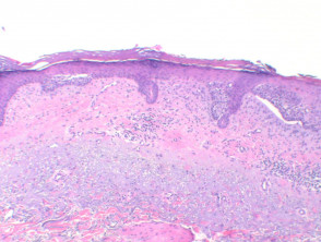
At low magnification, the distinctive feature of BCC is the presence of a tumor basaloid epithelium originating from the epidermis (Figure 1). This basaloid epithelium frequently organizes its cells forming a palisading pattern, which separates from the stroma underlying tumor by the formation of a clefting (Figure 2).
In the center of these tumor nests, crowded nuclei. nuclei, scattered mitotic mitotic figures, and evident apoptotic necrotic bodies (Figure 3) are observed. A crucial histological feature for differentiating it from other basaloid skin tumors is the presence of a mucinous stroma (Figure 4). Additionally, some tumors may show foci of regression, visible as areas of eosinophilic stroma where the basaloid nests are absent.
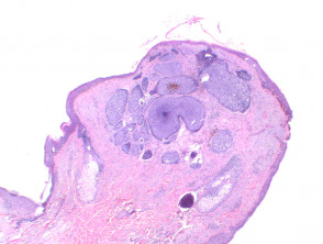
Figure 1
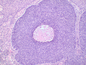
Figure 2
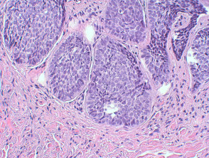
Figure 4
Staining and Special Histopathological Features
In immunohistochemical study, basal cell carcinoma generally stains positive for cytokeratin, although it tends to express cytokeratins more typical of infiltrate, thickening at the level of the. epithelium. While the BerEP4 marker is detected diffusely in the vast majority of these tumors, EMA expression is usually infrequent.
Common Variants of Basal Cell Carcinoma
Superficial BCC: This presentation is characterized by multiple focal nests of basaloid epithelium atypical. These nests emerge as buds from the basal layer. of the epidermis and tend to remain restricted to the level of the dermis papilla (Figure 5).
BCC Nodular: In this variant, the tumor shows formation of large masses or nests…
«`
presents a solid, growth pattern, manifesting as a nodule o nodules that has the capacity to invade surrounding tissues subcutaneous. Cartilage invasion is an uncommon finding (Figure 1).
Pigmented Basal Cell Carcinoma (Pigmented BCC): Focal deposits of melanin are observed distributed throughout the neoplasm. It is possible to identify a greater number of melanocytes within the tumor, and furthermore, there may be scattered and melanophages in the surrounding stroma (Figure 6).
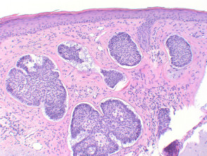
Figure 5
Special Stains and Syndromes Associated with Sebaceoma
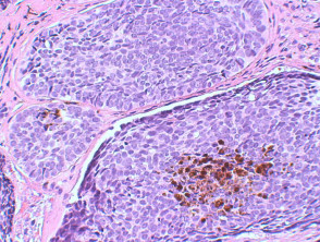
Figure 6
Micronodular BCC: Although it frequently presents a general nodular configuration, this tumor is histologically composed of multiple small nests. Characteristic of this subtype is its capacity to show extensive infiltration in the adjacent tissue, placing it among the subtypes with the prognosis least favorable prognosis, due to the higher risk of It is essential to remember that HNC exhibits a considerable rate of recurrence (Figures 7, 8, 9).
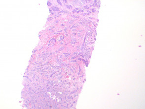
Figure 7
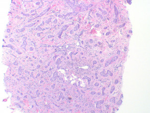
Figure 8
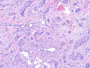
Figure 9
Basosquamous Carcinoma: Although it could be considered in the mucous membranes. of cell carcinoma cells, this tumor shares more clinical and histological similarities with basal cell carcinoma. The morphology cellular shows areas composed of pale-colored squamous cells, but they have the characteristic of lacking keratinization. keratinization. Therefore, it exhibits great analogy with the metatypical type. These tumors react positively to BerEP4 staining, while usually being negative for EMA.
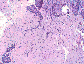
Infiltrative BCC: This subtype, categorized as aggressive, is distinguished by exhibiting extensive infiltrating strands.
y cords of atypical basaloid epithelium (Figures 10, 11).
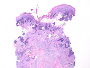
Figure 10
Pathology of Hemosiderotic Dermatofibroma
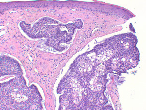
Figure 12
Metatypical BCC: This subtype, currently infrequent, illustrates the evolution of typical basal cell carcinoma towards areas characterized by large pale cells. In this variant, peripheral palisading and clefting are lost.
Infundibulocystic BCC: This variant is distinguished by the presence of multiple small cysts containing keratinous material, showing differentiation toward the infundibulum. follicular differentiation. Unlike trichoepithelioma, it lacks papillary mesenchymal bodies, although it often retains a mucinous stroma.
Sclerosing BCC: The tumor appearance in this form shows fine strands of atypical basaloid epithelium immersed in a dense and fibrotic.
Differentiation Sebaceous: Basal cell carcinoma can occasionally manifest significant areas of sebaceous differentiation (Figure 12). It is essential to differentiate this finding from sebaceoma, as will be detailed later.
Differential Diagnosis of Basal Cell Carcinoma
Trichoepithelioma: The epithelium of trichoepithelioma tends to integrate intimately with the surrounding matrix, which is frequently myxoid and fibrous. In contrast, basal cell carcinoma almost always exhibits some degree of retraction (clefting) between the epithelial cells and the adjacent tissue, potentially revealing mucin. stromal retraction. immunohistochemistry The presence of papillary mesenchymal bodies is a key indicator for trichoepithelioma. Generally,.
immunohistochemistryis not decisive, although staining with Bcl-2, Ber-Ep4, and CD34 are reported. BCL2 staining is usually diffusely positive in basal cell carcinoma, while it only delineates the basal layer in trichoepitheliomas. CD10 is positive in both basal carcinomas and trichoepitheliomas, although it tends to show specific peritumoral stromal reactivity.
Sebaceoma: This tumor frequently presents a more lobulated architecture and lacks the palisading, clefting, and mucinous stroma patterns characteristic of BCC. Immunostaining can be clarifying, as sebaceoma is rarely positive for BerEP4, while it is usually positive for EMA. desmoplastic. Microcystic Adnexal Carcinoma perineural : This diagnosis can be challenging when differentiating it from infiltrative basal cell carcinoma and trichoepithelioma glandular fever) variants. Merkel Cells Deep invasion, extensive.
stromal reaction, and convincing.


