
Understanding Atypical Mycobacterial Infections (NTM)
The atypical mycobacterial infections are caused by mycobacterial species other than Mycobacterium tuberculosis, the causative agent of pulmonary and extrapulmonary tuberculosis (including cutaneous TB), and organisms (, responsible for leprosy.
These nontuberculous mycobacteria (NTM) can trigger a wide range of human infections, which are fundamentally classified into four main clinical syndromes:
- Pulmonary disease.
- Histiocytic.
- Skin and soft tissue disease. soft tissues.
- Disease disseminated.
Specifically, atypical skin involvement manifests as the appearance of plaques y nodules crusted plaques and nodules. Additionally, in cases involving both the skin and bone structures, abscesses may develop. develop abscesses.
Clinical Manifestations of Cutaneous Infection by Atypical Mycobacteria
Etiology: Diversity of Species Causing Atypical Mycobacterial Infections
There is a wide diversity among mycobacterial species. Currently, at least 30 types of mycobacteria that are not responsible for either tuberculosis or leprosy have been identified. Among the most common NTMs that cause atypical mycobacterial infections are: atypical mycobacterial infections include:
- Mycobacterium avium-intracellulare
- Mycobacterium kansasii
- Mycobacterium marinum
- thrives best in environments with cooler temperatures, which explains its
- Mycobacterium chelonae
- Mycobacterium fortuitum
- Mycobacterium abscessus
While species such as Mycobacterium avium-intracellulare y Mycobacterium kansasii predominantly cause pulmonary disease that mimics TB, other strains such as Mycobacterium marinum, Mycobacterium ulcerans, Mycobacterium fortuitum y Mycobacterium chelonae are closely associated with the development of primarily cutaneous infections.
Clinical Manifestations of Atypical Mycobacterial Infections
The clinical manifestations associated with an atypical mycobacterial infection exhibit notable variability, which depends directly on the specific species of mycobacteria involved in the infectious process.
Clinical Presentation of the *Mycobacterium avium-intracellulare* Complex (MAC)
Infection caused by the *Mycobacterium avium* complex (MAC) is distinguished by a series of prominent systemic symptoms, especially in vulnerable populations:
- It constitutes the most prevalent nontuberculous mycobacterial (NTM) infection among patients diagnosed with AIDS.
- Systemic signs include persistent fevers, notable enlargement of the lymph nodes (lymphadenopathy), chronic diarrhea, debilitating fatigue, and significant, progressive weight loss.
- In some cases, this condition can progress to a form of pulmonary MAC infection.
- Associated dermal manifestations are infrequent and, when they appear, usually lack specificity.
Clinical Picture of *Mycobacterium kansasii* Infections
This species is the second most common cause of NTM infection in the context of HIV/AIDS, presenting clinical pictures with particular characteristics:
- It has the capacity to induce a chronic pulmonary infection that closely mimics the symptomatology of classic pulmonary tuberculosis.
- Frequent clinical signs include the appearance of fever, evident lymph node inflammation, and respiratory findings detectable on auscultation, such as crackles and wheezes.
- Cutaneous manifestations can be observed as single lesions or integrated within a more extensive disseminated disease.
Characteristics of *Mycobacterium marinum* Infections
*Mycobacterium marinum*, commonly known as fish tank or aquarium granuloma, is intrinsically linked to specific environmental exposure:
- It is a relatively rare infection, linked to recreational or occupational exposure to contaminated fresh or salt water sources.
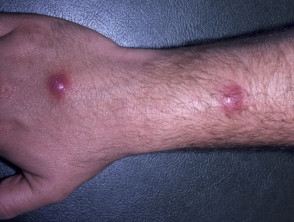
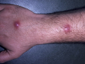
- Generally, the first clinical sign is the appearance of a solitary nodule or a pustule that, over time, can transform into a crusted sore or an abscess.
- It is common to observe the development of additional nodules following the path of lymphatic drainage, known as sporotrichoid presentation.
- The body regions most susceptible to involvement are commonly the elbows, knees, the dorsal surface of the feet, knuckles, or fingers.
- Immunocompromised individuals run a higher risk of developing multiple lesions and disseminated disease.
- Less frequently, it can manifest affecting structures near the inoculation site, causing joint symptoms such as bursitis, tenosynovitis, arthritis, or osteomyelitis, characterized by redness, noticeable inflammation, and localized pain in the affected joint.
Representative Images of *Mycobacterium marinum*
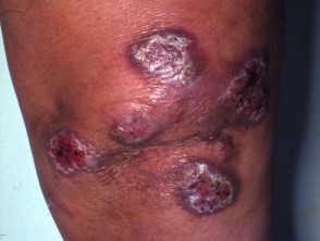
Mycobacterium marinum
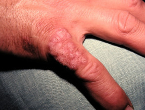
Mycobacterium marinum
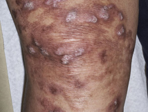
Mycobacterium marinum
Infections Caused by *Mycobacterium ulcerans*
*Mycobacterium ulcerans* is the etiologic agent responsible for conditions known as Buruli ulcer, Bairnsdale ulcer, or Kumasi ulcer. The infection presents the following epidemiological and clinical characteristics:
- Its prevalence is higher in Central and West African nations, particularly in environments with dense vegetation and swamps. However, cases have also been documented in regions like Australia.
- This microorganism is commonly found in aquatic environments, associated with fish and amphibians.
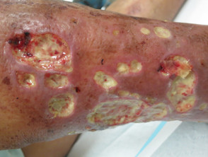
- After inoculation through a break in the skin barrier, an initial nodule nodule develops. This is painless, occasionally itchy, measures between 1 and 2 cm, and typically appears 7 to 14 days after infection.
- Over one to two months, this nodule can evolve into suppuration, originating a superficial ulcer that rapidly expands, compromising up to 15% of the patient's skin surface.
- The progression toward ulceration and ulceration and the necrosis tissue toxin destruction is induced by a specific toxin known as mycolactone.
- In cases of severe infection, there is a risk of destruction of blood vessels and nerves, blood vessels y nerves, with potential invasion up to the underlying bone tissue.
- A significant diagnostic challenge lies in the fact that the bacterial cultures required to isolate this microorganism grow extremely slowly.
Mycobacterium chelonae
- This infectious agent enjoys a wide distribution distribution, being a frequent contaminant in tap water and other water sources.
- Clinical manifestation can occur after trauma trauma involving tattoos or subsequent to surgical procedures.
- It primarily affects middle-aged individuals presenting with some level of Obesity..
- immunosuppression. It is capable of generating symptomatic conditions that include fever, pulmonary pathology, joint conditions, eye infections, and other forms of systemic infection.
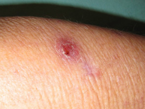
- Clinically, it can be observed as a chronic, hard-to-heal wound, a subcutaneous nodule subcutaneously, cellulitis, or the formation of localized abscesses.
- The existence of an immunosuppressed state can enhance the development of lesions that spread through various organs of the body.
Clinical Manifestations of Mycobacterium chelonae
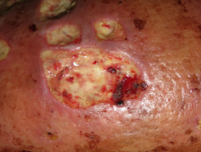
Mycobacterium chelonae
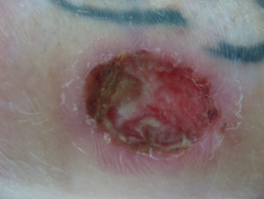
Mycobacterium chelonae
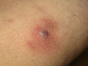
Mycobacterium chelonae
Mycobacterium abscessus
- This bacterium is formally classified as a subspecies belonging to the *M. chelonae* complex.
- Its natural reservoir includes water, soil, dust, and various animal species.
- Although it rarely compromises human health, when it does, it represents a considerable diagnostic and therapeutic challenge.
- It can cause dermal infections following puncture wounds, tattoos, recent skin trauma, or surgical interventions.
- It has the capacity to cause pulmonary infection and disseminated infections, especially in immunocompromised immunosuppressed.
Approach to Mycobacterium abscessus
Mycobacterium abscessus
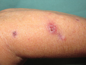
Mycobacterium abcessus
Mycobacterium fortuitum
- This bacillus also has an extensive global distribution, being detected in multiple environmental settings, including soil and bodies of water.
- It is isolated globally in both natural and treated water sources, and even in wastewater.
To obtain an accurate diagnosis and establish effective management for these atypical mycobacteria, it is essential to consider the patient's environmental exposure history and immune status.
- They present with lesions that have a predilection for extremities, elbows, knees, or genitals, and may be associated with previous septic foci, debris, or environmental contamination.
- Focal skin involvement, osteomyelitis, joint infections, or eye conditions eye drops may manifest after trauma (including tattoos or shaving followed by a foot bath).
- It commonly affects young individuals who are otherwise in good health.
- Profound immunosuppression, especially in AIDS patients, can precipitate the appearance of extensive skin and soft tissue lesions.
- Frequent sources of infection in wounds and surgical sites are often contaminated bodies of water.
- It causes the development of subcutaneous nodules and/or chronic ulcerative skin lesions that are difficult to heal, and furunculosis.
It is recommended to consult additional images regarding infections caused by atypical mycobacteria.
Diagnostic Procedures for Atypical Mycobacterial Infection
The definitive diagnosis of atypical mycobacteria is achieved through the culture culture of tissue. Because this process requires specific laboratory conditions, such as differentiated temperatures, it is crucial to inform staff about the existing clinical suspicion. Skin biopsy samples biopsy pathological findings lesions characteristic of these infections.
Other diagnostic methodologies employed include radiological studies and, more recently, the application of polymerase chain reaction (PCR) polymerase chain reaction (PCRtests on samples obtained from ulcers ulcers or via tissue biopsies biopsies.
Treatment Options for Atypical Mycobacterial Infection
The therapeutic regimen for atypical mycobacterial infections is adapted according to the specific organism organism responsible and the severity of the clinical picture. In most cases, antibiotic treatment is essential, involving drugs such as rifampin, ethambutol, isoniazid, minocycline, ciprofloxacin, clarithromycin, azithromycin, and cotrimoxazole. The standard protocol generally combines several of these agents.
When implementing antimicrobial therapy for atypical mycobacterial infections, it is important to consider the following specific details:
- Strains of Mycobacterium marinum Mycobacterium marinum antimicrobial are usually resistant to isoniazid, streptomycin, pyrazinamide, and para-aminosalicylic acid. Effective antimicrobial agents include tetracyclines, fluoroquinolones, macrolides (such as clarithromycin), rifampin, and sulfonamides (cotrimoxazole). The duration of treatment should be minimal, ranging from 4 to 6 weeks and potentially extending up to two months.
- The Candida infection Mycobacterium kansasii *Mycobacterium kansasii* requires a therapeutic regimen of at least three drugs maintained for 12 to 18 months. Rifampin is an indispensable pillar in this regimen.
- Mycobacterium chelonae y and respond best to clarithromycin or azithromycin when infections are localized, diseases, surgical debridement Once necrotizing fasciitis has been diagnosed, it is imperative to initiate therapeutic treatment immediately and aggressively. aminoglycoside aminoglycoside. (for example, combinations including amikacin, tobramycin, imipenem, and clarithromycin).
- The treatment for thrives best in environments with cooler temperatures, which explains its *Mycobacterium ulcerans* is most effective if initiated before lesions exceed 6 months of evolution and measure more than 10 cm in diameter. Currently recommended antibiotics are rifampin and streptomycin.
- Surgery is used as supportive therapy to antibiotic treatment in patients with severe infection. Although most lesions tend to heal spontaneously after six to nine months, it is common for them to leave significant scarring and disfigurement sequelae.
- In AIDS patients receiving therapy with HIV protease inhibitor drugs, and of SpeB, capable of dissolving tissue. of the HIV, rifampin should not be administered, as it significantly accelerates the breakdown of these antiretrovirals. In these circumstances, the use of rifabutin is the appropriate alternative.
In certain cases, it is necessary to proceed with the surgical removal of compromised lymph nodes and perform exhaustive debridement of the infected skin areas. Skin grafts may be required to repair surgical wounds resulting from severe cases.
Although some infections manage to resolve spontaneously, they will invariably produce a scar, scar, which is often unsightly.


