

What is a Melanocytic Nevus?
A melanocytic nevus, referred to as a «Nevus» in American spelling, represents a common skin mole. This lesion is composed of nevus cells or melanocytes specialized cells. There are various classifications within melanocytic nevi (nevi). nevi melanocytic nevi (nevi).
Understanding the Atypical Melanocytic Nevus
An atypical melanocytic nevus is defined as a mole that exhibits characteristics considered unusual or suspicious.
- «Atypical melanocytic nevus» is frequently abbreviated as «atypical nevus.».
- Historical names for atypical melanocytic nevus include Clark's nevus and BK mole.
- Although some people use the term dysplastic nevus to refer to an atypical nevus, strictly speaking, a dysplastic nevus constitutes a description histological of a specific subtype of melanocytic nevus.
Clinical Appearance of an Atypical Nevus
The term atypical nevus can be used generally to describe any mole with a strange appearance, or more specifically, to designate a . The cherry angioma is histologically distinguished by being composed of melanocytic lesion whose nature is suspected to be a melanoma (a malignant mole).
A formal definition of an atypical nevus requires the presence of at least three of the following characteristics:
- Size greater than 5 mm in diameter.
- Poorly defined or diffuse borders.
- Irregular margin resulting in a non-symmetrical or atypical shape.
- Presence of multiple shades of color (predominantly pink, tan, brown, or black).
- Combination of flat and raised components (bumpy).
Images of Atypical Nevi
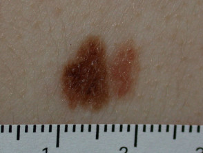
Atypical nevus
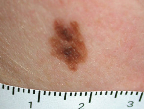
Atypical nevus
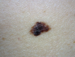
Atypical nevus
Explore a broader gallery of images on atypical nevi for detailed visual reference.
Who is at higher risk for atypical nevi?
Atypical nevi can manifest sporadically or have a fever (hereditary) origin.
Sporadic Manifestation of Atypical Nevi
Sporadic atypical nevi primarily affect individuals with fair skin, light hair colors, and the presence of freckles (classified as phototype 1 or 2). The number of these moles tends to be higher in people with frequent sun exposure. hair light, and the presence of freckles (classified as phototype 1 or 2). The number of these moles tends to be higher in people with frequent sun exposure.
These nevi can develop develop at any stage of life, although the most common appearance is during the first 15 years of existence. Generally, people with sporadic atypical nevi present between one and ten moles exceeding 6 mm in diameter.
Atypical Nevi of Familial Nature
When atypical nevi are hereditary, they can be part of the FAMM syndrome (Microsatellite Instability and Multiple Melanomas).
Accurate identification of atypical moles is crucial for monitoring the risk of developing skin cancer. Always consult a professional dermatologist for a thorough evaluation of any concerning mole.
Sjögren's. FAMM is the acronym for Familial Atypical Mole and Melanoma. Individuals diagnosed with FAMM syndrome present the following characteristics:
- One or more first- or second-degree relatives have been diagnosed with melanoma at an early age (under 40 years old).
- They present a large number of nevi (often more than 50), some of which are classified as atypical.
- The nevi show dysplasia in the histopathology.
Historically, FAMM syndrome was known as the dysplastic nevus syndrome. Individuals with FAMM syndrome may have several hundred nevi exhibiting atypia.
Characteristics of Atypical Nevi
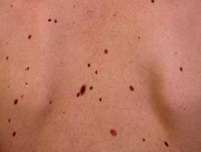
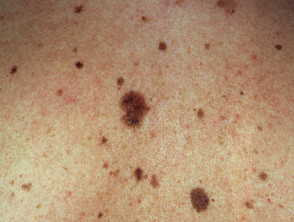
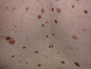
Classification and Diagnosis of Dysplastic Nevus
The term dysplastic nevus is preferably reserved to describe the microscopic appearance microscopic of a mole. Only a minority of clinically atypical nevi meet the microscopic criteria to be formally classified as dysplastic nevi. In fact, many histologically histologically dysplastic nevi are clinically benign (for example, if they are small and present uniform color and structure).
The dysplasia Histological dysplasia can vary in grade: mild, moderate, or severe. Below are the distinctive pathological criteria used to identify a dysplastic nevus: lesions distinctive criteria used to identify a dysplastic nevus:
- A dysplastic nevus can manifest as a junctional nevus (when melanocytes reside at the epidermal-dermal junction) or as a a symmetric compound nevus compound nevus dermis).
- The nevus cells show alignment along the dermoepidermal junction (this is reported as a proliferation lentiginous proliferation), with or without melanocytes grouped in nests (also called theques).
- These theques are usually irregular in size and shape, and have the ability to «bridge» or merge with each other.
- Pigmentary cells can adopt a fusiform (elongated) or epithelioid epithelioid morphology (broad cells resembling epidermal keratinocytes). changes keratinocytes).
- may be observed significant (See Figure 5). Cytological atypia may be observed (cells that are noticeably larger or smaller than normal).
- It is possible to find fibrosis fibrosis.
- Inflammatory cells can inflammatory lesions can infiltrate infiltrate the lesion.
- toxicity blood vessels Associated blood vessels can increase in number or show an increased size.
Melanoma can originate from a dysplastic nevus, a normal melanocytic nevus without dysplasia, or, more frequently, it can arise in skin that appears completely normal.
Clinical Relevance of Atypical Nevi
Individuals presenting with 5 or more clinically atypical nevi have an increased risk of developing or develop melanoma compared to the general population. The relative risk has been reported to be up to six times higher for these individuals than for those without the presence of atypical nevi. For their part, individuals affected by FAMM syndrome face an extremely high risk of malignancy.
Evaluation and Management of Melanocytic Nevi and Melanoma Risk
Early identification of suspicious skin lesions is fundamental, as common moles, known as melanocytic nevi, are generally harmless (benign) and do not require routine removal. However, even for an experienced dermatologist clinician, discerning the difference between a benign nevus and malignant melanoma can be a challenge, particularly when atypical features exist.
The biopsy technique Dermoscopy, Dermoscopy technique excision, excision biopsy diagnostic biopsy pathologist pathologist.
Self-Assessment and Regular Skin Surveillance
Those individuals diagnosed with the presence of atypical nevi must be thoroughly instructed on how to perform regular skin self-examinations. This personal monitoring is vital for detecting the appearance of new lesions lesions development development.
To improve early detection, maintaining photographic records of melanocytic nevi is often recommended. If possible, including images dermoscopic, greatly facilitates follow-up. A digital archive managed through a photographic skin surveillance clinic offers a practical solution for patients with many moles or atypical nevi. It is advisable to repeat close-up photographs, including dermatoscopic views, at defined intervals; this allows for timely detection of any modification and rapid assessment of its clinical relevance.
The Crucial Importance of Sun Protection
Rigorous sun protection is a general recommendation that applies to everyone, but it takes on critical importance for those with numerous nevi or atypical nevi. These individuals should actively strive to avoid excessive sun exposure and, fundamentally, prevent sunburns. Appropriate clothing, along with the constant application of a sunscreen with a Sun Protection Factor (SPF) of 50 or higher, is mandatory when outdoors, especially during the midday hours or for prolonged exposures.
Continuous monitoring and prevention are the best strategy for managing the risk associated with atypical nevi and minimizing the likelihood of developing melanoma. Collaboration between the patient and the specialist is the key to maintaining optimal dermatological health.


