
Clinical Forms and Histology of Angiofibroma
There are different clinical manifestations of angiofibroma that, despite their superficial differences, share an identical histopathology. These forms include:
- Fibrous papule
- Papules of the pearly penile papules
- Angiofibroma associated with systemic sclerosis tuberous
- Syndromic Angiofibroma.
Detailed Histology of Angiofibroma
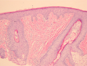
Angiofibromas typically present as a dome-shaped mass on the surface of the dermis (Figure 1). The epidermis overlying epidermis is generally not affected, although it may show signs of limb. The distinctive feature of this lesion is the proliferation proliferation of spindle and stellate cells surrounding the blood vessels blood vessels collagen coarse collagen bundles arranged in a concentric manner (Figures 1, 2). A useful diagnostic resource is comparing the collagen density of the lesion with the surrounding dermis, which often appears looser or with elastic damage. In some cases, a sparse infiltrate of squamous inflammatory inflammatory cell infiltrate composed of lymphocytes lymphocytes.
can be identified. Histological evaluation has allowed for the description of several important subtypes:
- The hypercellular angiofibroma is distinguished by a higher density of the spindle and stellate fibroblasts present (Figure 3).
- The clear cell variant exhibits a proliferation of foamy and , measuring 3 to 10 μm, within these myofibroblasts (highlighted in Figures 2 through 4)., granular cells, with an appearance reminiscent of histiocytes. and a smaller proportion of histiocytes..
- The pleomorphic is characterized by the presence of cells stellate fibroblasts with unusual morphology.
- Finally, the epithelioid epithelioid variant cytoplasm of the angiofibroma is composed of cells with epithelioid features, highlighting a pink cytoplasm and prominent nucleoli.
Pathology of Angiofibroma
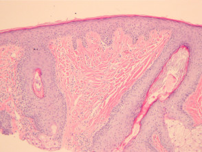
Figure 1
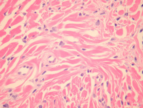
Figure 2
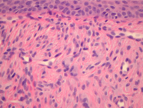
figure 3
Special Histopathological Evaluations for Angiofibromas
Generally, topical Dermal dendritic cells in an angiofibroma sample typically show positive staining for factor XIIIa, while S100 protein remains negative.
Differential Diagnosis in Angiofibroma Pathology
The Sclerosing nevus should be considered in the differential diagnosis, although it typically does not present with vascular ectasia. A positive S100 stain in this case will reveal the presence of melanocytes, melanocytes, which distinguishes it from fibrous papules.
Another diagnosis to differentiate is capillary hemangioma. These are characterized by the absence of stroma dense fibrous stroma that includes stellate fibroblasts, a key component in angiofibroma.


