

Acral Lentiginous Melanoma: What It Is, Who Gets It, and How to Identify It
Understanding Acral Lentiginous Melanoma
Acral lentiginous melanoma is a specific form of melanoma that appears on the palms of the hands, the soles of the feet, or under the nail (subungual melanoma). Its defining characteristic is its anatomical location: it affects the palmoplantar areas. Generally, it is more prevalent on the feet than on the hands. This lesion can originate de novo on normal-looking skin, or it can evolve from a pre-existing melanocytic nevus (mole).
Initially, acral lentiginous melanoma presents as a flat patch that grows slowly. During this initial stage, the malignant cells remain confined to the tissue of origin, the epidermis. This is the stage in situ of melanoma, which can persist for several months or even years without invading deeper layers.
The transition to an invasive stage occurs when cancer cells cross the basal membrane of the epidermis and enter the dermis. Additionally, rapid nodular growth can also develop secondarily within an established acral lentiginous melanoma, proliferating deeper into the skin structure.
Who is Most Prone to Acral Lentiginous Melanoma?
In countries like Australia and New Zealand, acral lentiginous melanoma accounts for between 1% and 3% of all melanoma cases. Melanoma is a potentially serious skin cancer derived from pigment cells, known as melanocytes. While it is a less frequent subtype in fair-skinned people, it constitutes the most common variant of melanoma in individuals with darker skin tones.
Compared to other types of melanoma, acral lentiginous is relatively rare. Curiously, its incidence does not seem to be directly linked to skin phototype; it occurs at similar rates in light, olive, or black skin. However, while it accounts for between 29% and 72% of melanomas in dark-skinned individuals, in fair-skinned people it is responsible for less than 1% of melanomas. This is because fair-skinned people are more susceptible to sun-induced melanoma types, such as superficial spreading melanoma and lentigo maligna melanoma.
Diagnosis occurs with equal frequency in both men and women, predominantly affecting people over 40 years of age. To date, the specific cause of acral lentiginous melanoma is still unknown, and a direct relationship with sun exposure has not been established.
Visual Aspects of Acral Lentiginous Melanoma
When acral lentiginous melanoma appears on the palm, sole, or subungual area, it initially manifests as an expanded patch of skin with altered pigmentation. Frequently, at the beginning, it is confused with a simple freckle or spot by the patient. It can also present as an amelanotic lesion (without discernible pigmentation, often red).
When forming a flat skin lesion, its signs can be evaluated using the ABCDE mnemonic rule, common to all melanomas: Asymmetry, Border irregularity, Color variation, large Diameter, and Evolution or change over time.
The distinctive characteristics of acral lentiginous melanoma include:
- Considerable size: It tends to be larger than 6 mm, often reaching several centimeters in diameter at the time of diagnosis.
- Variable pigmentation: It commonly exhibits a mix of tones, including brown, grayish-blue,
Early detection is crucial to improve the prognosis of any type of skin cancer. If you notice persistent changes in moles or suspicious appearances on palms, soles, or under the nails, consult a dermatologist immediately for a professional evaluation.
- A color that varies between brown, black, and red.
- A surface that may initially be smooth, evolving into a thicker, irregular, and possibly dry or warty.
- Possibility of infection ulceration or bleeding.
Acral Lentiginous Melanoma
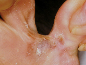
Acral Lentiginous Melanoma
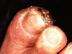
Acral Lentiginous Melanoma
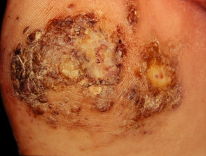
Acral Lentiginous Melanoma
Explore more relevant images about acral lentiginous melanoma for better visual understanding.
Subungual Melanoma
When acral lentiginous melanoma is located in the nail area, it is called nail unit melanoma. If its origin is the matrix (where the nail grows), it is specifically classified as subungual melanoma. Clinically, it can manifest as generalized discoloration or as one or more longitudinal pigmented bands longitudinal over the The nail plate. In advanced stages, this melanoma can completely erode the nail plate and, occasionally, may present without pigmentation (amelanotic).
Nail Unit Melanoma
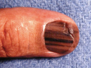
Subungual melanoma
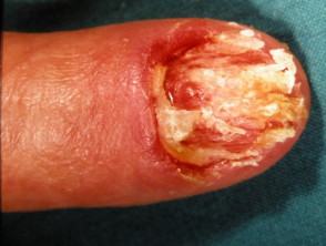
Subungual melanoma
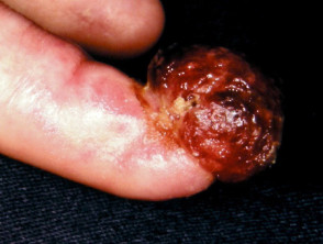
Subungual melanoma
See additional images about nail unit melanoma.
What is the Cause of Acral Lentiginous Melanoma?
Acral lentiginous melanoma originates from the malignant transformation of pigment cells (melanocytes) that proliferate along the basal layer. layer of the epidermis. These cancer cells can develop from a pre-existing melanocytic nevus or, more frequently, on skin with a normal appearance. Although the exact trigger that induces the malignant transformation of melanocytes is unknown, it is known that this process is intimately related to mutations are being investigated..
Diagnostic Tests for Acral Lentiginous Melanoma
Dermoscopy
The A positive result could indicate that the patient requires periodic exams for other types of cancer. For example, a patient with a mutation in the, Dermoscopy asymptomatic by a dermatologist dermatologist or other medical professional with appropriate training, is an extremely valuable diagnostic tool for differentiating acral lentiginous melanoma from other skin conditions.
Diagnosis and Characteristics of Acral Lentiginous Melanoma: Biopsy and Pathology Reports
Other lesions Other skin lesions, particularly:
- Nevi Melanocytic Nevi (moles)
- Viral warts
- Hemorrhage: subungual The clinical presentation of aneurysmal dermatofibroma manifests as a bluish-brown nodule that undergoes rapid growth. This accelerated growth is secondary to episodes of o hematoma subungual space
The most frequently observed dermoscopic features in acral lentiginous melanoma are:
- Asymmetrical structure and coloration asymmetrical
- Parallel distribution pattern
- Grayish-blue structures.
Consult the section on dermoscopy of acral lentiginous melanoma for more details.
Skin of Acral Lentiginous Melanoma
If acral lentiginous melanoma is suspected in a . The cherry angioma is histologically distinguished by being composed of lesion, the best practice is complete excision Excision. Partial biopsy should be avoided unless lesions are exceptionally large. An incisional or punch biopsy runs the risk of omitting a focus of melanoma that might be emerging within a pre-existing nevus. However, in cases of very large lesions, a preliminary biopsy can be arranged to confirm the diagnosis before major surgery. The biopsy of a lesion suspicious for acral lentiginous melanoma should remove a long ellipse of skin, or multiple incisional biopsies biopsies from various sites should be obtained, as a single spot might not capture a malignant focus.
Actinic keratosis. pathological Pathological diagnosis histologic histological features proliferation asymmetrical proliferation of melanocytes at the dermoepidermal junction.
Key Elements of the Pathology Report Pathology
Pathology Report Details pathologist The pathologist's report macroscopic macroscopic description microscopic detailed microscopic description, covering the following crucial points:
- Diagnosis of primary melanoma primary
- Breslow Thickness, Breslow thickness, measured to the nearest 0.1 mm
- Clark invasion level
- Excision margins: Evaluation of the normal tissue surrounding the tumor
- Clark invasion level. mitoticMitotic rate proliferate
- Presence or absence of ulceration
Additionally, the report may contain observations on cell type, growth pattern, invasion into blood vessels o nerves, : a metric that measures the rate at which cells are inflammatory, blood vessels or nerves, inflammatory response, regression and whether an associated nevus (the original mole) exists.
What is Breslow Thickness?
The Breslow thickness is determined for invasive melanomas. This measurement is taken vertically in millimeters, from the surface of the granular layer granulomatous skin layer Benefits of Germline Genetic Testing for Melanoma metastases.
What is the Clark Invasion Level?
The Clark level classifies the specific anatomical plane where tumor invasion has occurred:
Level 1: Melanoma in situ
papillary dermis papillary dermis
Level 3: Melanoma has completely occupied the papillary dermis
reticular dermis reticular
subcutaneous tissue subcutaneously
metastases Benefits of Germline Genetic Testing for Melanoma A deeper Clark level indicates a higher risk of secondary metastases. This system is particularly useful for predicting prognosis in thin tumors, whereas for thicker tumors, Breslow thickness is usually considered a more robust prognostic indicator.
Treatment of Acral Lentiginous Melanoma
The initial treatment for primary melanoma always consists of its removal;
The lesion must be completely excised, ensuring a margin of 2 to 3 mm of healthy tissue around it. Subsequent treatment is primarily defined by the Breslow thickness of the excised lesion.
Following initial excision biopsy, the Australian and New Zealand guidelines for melanoma treatment (2008) establish recommended radial excision margins, measured from the clinical edge of the melanoma, as detailed in the following table. It is important to note that wound closure may require a flap or a graft. In the specific case of acral and subungual lentiginous melanoma, treatment might involve partial amputation of a finger. digit radiotherapy radiotherapy.
Melanoma in situ - 5 mm excision margin
Melanoma <1.0 mm - 1 cm excision margin
Melanoma 1.0–2.0 mm - 1–2 cm excision margin
Melanoma 2.0–4.0 mm - 1–2 cm excision margin
Melanoma > 4.0 mm - 2 cm excision margin
Melanoma Staging and Prognosis
Melanoma staging is the crucial process for determining if the cancer has spread from its primary location on the skin. Most specialists are guided by the staging guidelines for cutaneous melanoma cutaneous from the American Joint Committee on Cancer (AJCC) of 2009. Generally speaking, the stages are classified as follows:
- Stage 0: Melanoma in situ
- Stage 1: Thin melanoma (less than 2 mm thick)
- Stage 2: Thick melanoma (greater than 2 mm thick)
- lymph nodes lymph nodes
- Stage 4: Detection of Benefits of Germline Genetic Testing for Melanoma distant metastases
Melanoma > 2 mm: 20 mm.
If local lymph nodes are inflamed due to metastatic melanoma, metastatic, general anesthesia over-the-counter General anesthesia. If the nodes are not visibly enlarged, they can be examined to rule out microscopic spread of melanoma through a test called sentinel lymph node biopsy.
In New Zealand, many surgeons suggest sentinel lymph node biopsy for melanomas thicker than 1 mm, especially in younger patients. Nevertheless, although this biopsy aids in cancer staging, it has not currently shown a direct survival advantage, so its necessity remains a topic of clinical debate.
Lymph nodes harboring metastatic melanoma often increase rapidly in size. Typically, an affected node is not tender to the touch and presents a firm to hard consistency. If you notice this change between scheduled follow-up appointments, it is crucial to notify your doctor immediately.
If the melanoma has widespread, spread and of BRAF BRAF inhibitors.
Post-Treatment Follow-up Protocol
The fundamental purpose of follow-up is to identify any recurrence of the disease in its initial phases.
The Guidelines for the Management of Melanoma in Australia and New Zealand (2008) suggest the following guidelines for monitoring patients with invasive melanoma:
- Perform regular skin self-examinations.
- Schedule routine skin checks with the patient's preferred healthcare professional.
- Follow-up intervals should be every six months for the first five years for patients with Stage 1 disease. For Stages 2 or 3, quarterly or every four months checks are recommended, transitioning to annual check-ups after that period for all patients.
- It is vital to evaluate the individual needs of the patient before establishing an appropriate follow-up plan.
- Provide comprehensive education and support to facilitate the patient's adaptation to their condition.
Follow-up appointments can be managed by the primary care physician, the treating specialist, or through a shared care model.
Follow-up checks may include evaluations such as:
- Examination of the scar scar Individual needs should be considered before determining an appropriate follow-up schedule.
- Palpation of regional lymph nodes to detect abnormalities.
For those patients with more advanced primary disease, follow-up may be expanded to include:
- Blood tests, including LDH measurement.
- Advanced imaging techniques: ultrasound: This tool is useful for diagnosing chronic, ultrasound CT (Computed Tomography): This scan shows exclusive skin thickening in cases of lipedema. In lymphedema, in contrast, fluid accumulation, a characteristic honeycomb pattern, and possible muscle enlargement are observed., Magnetic Resonance Imaging (MRI) CT scans, MRI, and PET scan. PET.
recurrence It is essential to remember that HNC exhibits a considerable rate of or metastasis. Furthermore, it is not considered necessary to monitor healthy patients who have remained disease-free for five years or more after melanoma excision, regardless of their initial stage, with tests.
Long-Term Outlook for Melanoma
Acral lentiginous melanoma that remains in situ in situ.
Melanoma guidelines indicate that metastases are infrequent in melanomas with a thickness under 0.75 mm, and the risk is around 5% for tumors between 0.75 and 1 mm thick. The risk progressively increases with thickness; for example, melanomas over 4 mm have a 10-year survival rate of approximately 50%, according to statistics provided by the American Joint Committee on Cancer (AJCC).
Proper management of the primary lesion and rigorous follow-up are fundamental to ensuring the longest recurrence-free survival time. Continue to collaborate closely with your medical team to adhere to the personalized monitoring protocol.


