
Understanding Epidermolysis Bullosa and Its Acquired Variant
The Epidermolysis bullosa (EB) represents a group of hereditary conditions characterized by blister formation, which are present from birth.
What is Epidermolysis Bullosa Acquisita (EBA)?
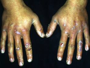
The Epidermolysis bullosa acquisita (EBA) is a disease defect that is rare. It manifests with the appearance of subepidermal tense blisters located specifically in areas subject to trauma physical trauma. A key differentiating factor is that, unlike EB, EBA is not inherited and usually manifests during adulthood.
EBA blisters tend to concentrate in areas that are easily damaged, such as the hands, feet, knees, elbows, and the gluteal region. Occasionally, there may be mucosal mucosa, involvement, causing blister formation in the oral cavity, nose, and eyes.
Illustrative Images of Epidermolysis Bullosa Acquisita
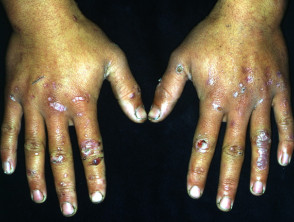
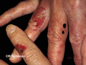
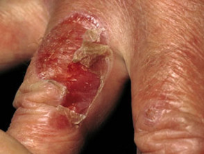
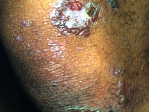
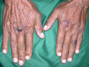
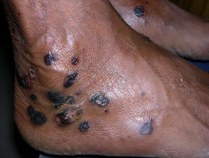
It is essential to understand that while EB is a genetic condition present from birth, Epidermolysis bullosa acquisita is an autoimmune entity that requires a different diagnostic and therapeutic approach than when facing hereditary forms. The localization of damage in friction sites is a key indicator for suspecting EBA in adults.
Who Develops Epidermolysis Bullosa Acquisita (EBA) and Risk Factors?
Epidermolysis bullosa acquisita (EBA) generally manifests in the fourth and fifth decades of patients' lives. It affects both men and women of any ethnic background.
In some individuals with EBA, the coexistence of other autoimmune conditions has been observed, with Crohn's disease and systemic lupus erythematosus being the most frequent associations. Health problems such as amyloidosis, multiple myeloma, and, rarely, lung carcinoma and lymphoma are also reported. However, other patients only present with skin involvement without these comorbidities.
Understanding the Etiology of Epidermolysis Bullosa Acquisita
Researchers have identified the presence of Immunoglobulin G (IgG) autoantibodies directed against the NC1 domain of type VII collagen (Col7) in affected patients. Type VII collagen is a fundamental structural component of anchoring fibrils. These fibrils connect the epidermal basement membrane to the underlying dermis.
The destruction of these anchoring fibrils results in blister formation just below the epidermis, in a region called the lamina densa. This autoimmune process is the central mechanism that triggers EBA pathology.
Distinctive Clinical Presentations of Epidermolysis Bullosa Acquisita
EBA manifests through several well-differentiated clinical presentations, each with specific patterns of involvement and prognosis.
| EBA Classification | Key Clinical Features |
|---|---|
|
Classic Mechanobullous Form of EBA |
|
|
Non-Classic or Non-Mechanobullous Form of EBA |
|
|
Predominantly Mucous Membrane Form of EBA |
|
|
Brunsting-Perry EBA Type |
|
|
IgA-EBA |
|
Diagnosis of Epidermolysis Bullosa Acquisita (EBA)
The definitive diagnosis of Epidermolysis Bullosa Acquisita (EBA) requires performing several specialized clinical and laboratory tests to confirm autoimmunity and characteristic dermal involvement.
Key Tests for EBA Diagnosis
To establish an accurate diagnosis of EBA, the following main analyses are required:
- Skin Biopsy: This histopathological procedure will reveal the presence of a subepidermal blister, which indicates the separation between the epidermis and the dermis. Cells of a inflammatory infiltrate mixed inflammatory infiltrate are also observed in the dermis, and milia cysts and scars are common in older lesions.
- Direct Immunofluorescence (DIF) of Skin Biopsy: DIF analysis typically shows the linear deposition of Immunoglobulin G (IgG, more frequent than IgM or IgA) and C3 along the dermal-epidermal junction. It is important to note that blood blood vessels vessels are not usually involved in this deposition pattern.
- Indirect Immunofluorescence (IIF) of Serum: This test detects the presence of circulating IgG autoantibodies, generally at a low titer, which are specifically directed against the basement membrane.
There are additional tests only available at specialized academic centers for exhaustive confirmation of the diagnosis [2]:
- Skin biopsy for advanced studies such as direct and indirect immuno-electron microscopy, microscopy, analysis of the salt-split skin DIF pattern, antigenic mapping by fluorescence overlay, and salt-split skin DIF.
- Blood analysis to detect undetectable circulating antibodies specific against Type VII Collagen (Col7), using techniques such as immunoblotting, enzyme-linked immunosorbent assay (ELISA), more detailed IIF, and immunoprecipitation.
The International Bullous Diseases Group (IBDG) has established consensus diagnostic criteria for EBA, which are crucial for confirmation [1,2]. If the clinical and histological presentation of the blistering disease is compatible with EBA, the diagnosis is considered confirmed if two main conditions are met: histological of the blistering disease is compatible with EBA, the diagnosis is considered confirmed if two main conditions are met:
- Demonstration of linear deposition of IgG and/or C3 along the basement membrane zone by direct immunofluorescence (DIF).
- Detection of anti-COL7 autoantibodies in the patient's serum (by enzyme-linked immunosorbent assay).
Differential Diagnosis of Epidermolysis Bullosa Acquisita
EBA presents a diagnostic challenge, as it can mimic the clinical appearance of other inflammatory blistering diseases. The conditions most frequently considered in the differential diagnosis include bullous pemphigoid and bullous systemic lupus erythematosus (the latter also associated with anti-Col7 autoantibodies).
When contrasting EBA with bullous pemphigoid, EBA is distinguished by several key features [2]:
- It tends to manifest in younger individuals, generally under 70 years of age.
- It presents prominent involvement of the head and neck.
- It frequently involves the mucous membranes.
- Skin lesions tend to heal leaving noticeable scars and milia formation.
Treatment Options for Epidermolysis Bullosa Acquisita
The The primary in the management of EBA is twofold: protecting the skin to stop the formation of new blisters and simultaneously promoting the healing of existing lesions to prevent long-term complications.
Since EBA is classified as an autoimmune disease, the use of immunosuppressive immunosuppressive agents
- A biopsy may reveal deposits of
- Dapsone
- Colchicine
- Corticosteroids
- Mycophenolate Mofetil
- Images of Pigmentary Lichen Planus
- Intravenous immunoglobulin.
Due to the rarity of this condition, establishing with certainty which of these drugs offers maximum efficacy remains a clinical challenge.
In addition to immunosuppressive treatment, there are other fundamental management strategies for the patient's quality of life:
- Proactive treatment of any underlying systemic disease.
- Coordination and consultation with specialists such as dentists and ophthalmologists, ophthalmologists, depending on the location of the lesions.
- Implementing strict measures to avoid direct physical trauma to vulnerable skin surfaces.
- In patients with oral involvement, advice should be given to avoid consuming excessively hard, brittle, or highly acidic foods.
Prognosis of Epidermolysis Bullosa Acquisita
EBA is characterized as a chronic inflammatory dermatosis that proceeds in cycles of remissions partial remissions and exacerbations. exacerbations. With appropriate treatment and rigorous care, patients have the expectation of leading a normal life. However, there are potential risks to manage, including:
- Risk of secondary Risk of infection.
- Development of severe scars that can lead to joint physical deformities and limitation of range of motion.
- Periodontal complications.
- Risk of conjunctival scarring that can progress to blindness.
- Possible side effects from long-term immunosuppressive medication.
Comprehensive management and control of EBA-specific autoantibodies are essential to positively influence the long-term prognosis of patients affected by this rare disease.


