
Toxic epidermal Necrolysis (TEN) is a severe manifestation of cutaneous drug reactions. It begins with flu-like symptoms (prodromal phase) and rapidly progresses to painful, widespread erythematous rash, followed by extensive sloughing of the skin and mucosal surfaces. TEN occupies the most critical end of the spectrum, contrasting with Stevens-Johnson Syndrome, because TEN involves detachment exceeding 30% of the total body surface area.
Histopathology of Toxic Epidermal Necrolysis: Key Differentiating Features and Diagnosis
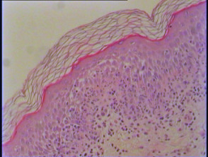
Histopathologically, TEN is characterized by the development of subepidermal bullae (illustrated in Figures 1–3). These structures are linked to extensive epidermal necrosis, leading to the detachment or complete eradication of the epidermis. At the advancing edge of this necrosis, apoptotic keratinocytes can frequently be identified (Figure 2, arrow). A crucial diagnostic feature of TEN is the unexpectedly minimal underlying inflammatory infiltrate observed in the dermis.
Pathology Findings Illustrating Toxic Epidermal Necrolysis
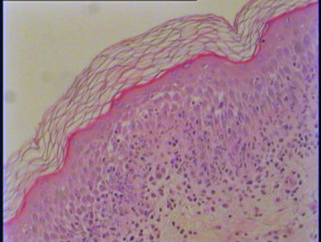
Figure 1
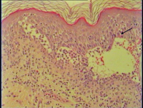
Figure 2
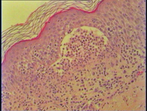
Figure 3
Establishing the Differential Diagnosis for Toxic Epidermal Necrolysis
Erythema multiforme presents differently; while it involves apoptotic keratinocytes, the necrosis tends to be less extensive (often central to the lesion). Furthermore, the associated dermal inflammation is generally more prominent, frequently featuring a lymphocytic perivascular infiltrate.
Regarding Phototoxic Reactions, these also manifest with significant edema and subepidermal blistering, alongside necrotic keratinocytes and minimal infiltrate. However, careful clinical context and history are usually sufficient to separate these from true TEN.
In cases of severe Chemotherapy-induced acral erythema, subepidermal bullae and epidermal necrosis can develop. Despite these similarities in tissue damage, this condition possesses distinct clinical markers differentiating it from TEN.
The comprehensive differential diagnosis for conditions exhibiting subepidermal blistering concurrent with sparse cell death must also incorporate epidermolysis bullosa variants, porphyria cutanea tarda, cell-poor bullous pemphigoid, acute thermal burns, suction-induced blisters, and other specific drug-induced bullous disorders.
Accurate differentiation of TEN from these similar-appearing dermatoses is critical for initiating appropriate, life-saving dermatologic care promptly.


