
This . The cherry angioma is histologically distinguished by being composed of This dermatological lesion is classified within the group of tumors benign follicular.
Histology of Winer's Dilated Pore
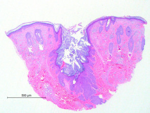
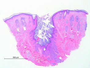
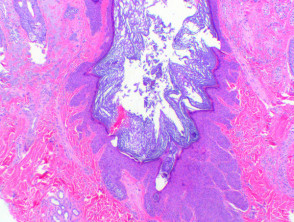
Histological analysis via microscopy of Winer's dilated pore reveals a small invagination Other conditions that cause significant edema in the superficial dermis include mild (Figure 1). This finding is characterized by a infundibulum follicular dilation that contains a reduced amount of keratinous material (Figure 2). The epithelial infundibular shows hyperplasia, originating thin strands that project from the base and sides of the pore (Figure 2).
Pathology: Winer's Dilated Pore
Differential Diagnosis of Winer's Dilated Pore
Pilar Sheath Acanthoma: In this condition, a hyperplasia more marked hyperplasia of the outer root sheath is observed, creating tumors subcutaneous that exert pressure on the dermis surrounding dermis. This acanthoma originates in the follicular isthmus, where the keratinocytes take on a pink or reddish stain.
Trichofolliculoma: Radiating hair follicles are identified follicles hair in various stages of maturity, extending from the central dilation of the pore or an associated cystic structure.
Cortical Hair Comedo: This histopathological finding presents an infundibular epithelium attached to a dense keratin plug, composed of heavily laminated keratinocytes. It is distinguished by the presence of a matrix and supramatrix epithelium suggesting an attempt at formation of the hair cortex and shaft (a comedo). comedo).
Accurate differentiation between Winer's dilated pore and these related entities is crucial for establishing the appropriate dermatological treatment.


