
«`html
Warfarin Necrosis: Understanding Paradoxical Coagulation
Warfarin necrosis is a rare paradoxical complication in patients undergoing anticoagulant therapy with warfarin. It occurs in approximately one in every 10,000 patients prescribed this medication, marking an unexpected adverse response.
Histopathology of Warfarin-Induced Necrosis
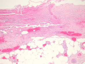
Histological analysis of warfarin necrosis reveals variable degrees of necrotic damage in both the epidermis and the dermis (Figure 1). Extensive intravascular thrombi are observed within capillaries and venules (Figures 2 and 3). The remaining patent vessels are dilated, reflecting microvascular obstruction.
Pathological Findings of Warfarin Necrosis
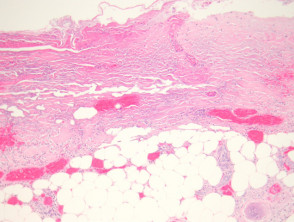
Figure 1
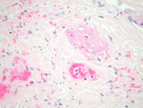
Figure 2
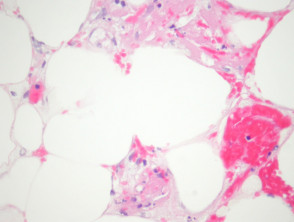
figure 3
Diagnostic Evaluation of Warfarin Necrosis
In most cases of suspected warfarin necrosis, additional specialized histological or laboratory studies are not required to confirm the diagnosis.
Differential Diagnosis of Warfarin Reaction
The differential diagnosis of warfarin necrosis requires distinguishing this entity from other thrombotic skin pathologies. It is crucial to differentiate it from disseminated intravascular coagulation (DIC) or any other coagulopathy, as they can be morphologically identical. Therefore, direct correlation with the patient's clinical history is essential.
Likewise, one must consider septic thrombi. In these cases, the presence of neutrophils and other inflammatory cells is generally identified. The use of special stains to detect microorganisms, along with correlation with blood culture results, can be useful in establishing a definitive diagnosis and ruling out an infectious etiology.
```


