
Histology and Microscopic Characteristics of Tinea Versicolor
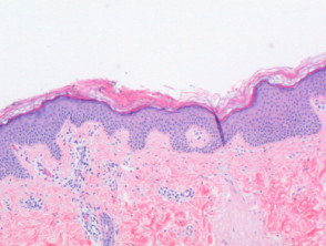
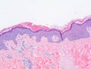
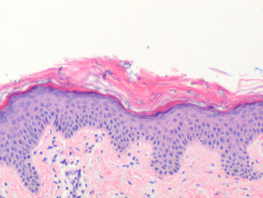
When examining tinea versicolor at low power in histology, a mild perivascular inflammation with a characteristic pattern is suggested (Figure 1). In the epithelial tissue, the epidermis may show signs of mild hyperkeratosis and acanthosis (Figure 2). It is also possible to observe an elongation of the rete ridges and a slight increase in basal layer pigmentation, findings that sometimes resemble those seen in acanthosis nigricans.
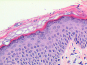
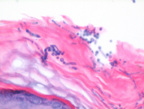
Mainly, the microorganisms causing tinea versicolor are located almost exclusively within the stratum corneum, facilitating their visualization in hematoxylin and eosin stained sections (Figures 2 and 3). The distinctive feature is the short hyphae and spores of *Malassezia furfur*, often described with the analogy of «spaghetti and meatballs» or «cigar butts» (Figure 4). Additionally, a mild superficial perivascular lymphocytic infiltrate may be detected, occasionally accompanied by plasma cells.
Key Findings in the Pathology of Tinea Versicolor
Improving Diagnosis with Special Stains in Tinea Versicolor
The use of specialized stains, such as periodic acid-Schiff (PAS) and silver methenamine, is essential to improve the visualization and confirmation of fungal organisms present within the stratum corneum.
Differential Diagnosis of Tinea Versicolor
Although the identification of characteristic fungal forms in the appropriate clinical context is usually sufficient for diagnosis, in cases where fungal findings are scarce, it is crucial to consider the following list of differential diagnoses:
- Gougerot and Carteaud Confluent and Reticulated Papillomatosis: Although similar epidermal alterations may be observed, in this condition the degree of hyperkeratosis and downward acanthotic growth tend to be more marked. Normally, very few or no fungal elements are identified.
- Acanthosis Nigricans: In exceptional cases, and when not linked to underlying pathology, spore forms may be observed in the stratum corneum.


