
Tinea Manuum: Definition, Clinical Features, and Risk Factors
The tinea manuum is defined as a secondary fungal infection caused by dermatophytes affecting one or both hands. This condition is significantly less common than tinea pedis (fungal infection of the feet).
Tinea manuum is often misdiagnosed due to its clinical resemblance to other skin conditions, such as:
- Hand dermatitis, specifically the variant known as pompholyx.
- Psoriasis, particularly palmoplantar pustular.
- Exfoliative keratolysis.
Images of Tinea Manuum
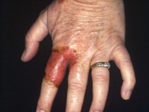
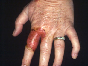
Tinea Manuum
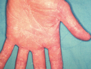
tinea manuum
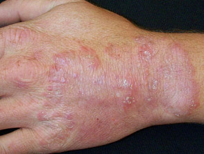
tinea manuum
Distinctive Clinical Features of Tinea Manuum
The presentation of tinea manuum can vary considerably. In some cases, it appears as an acute, inflammatory rash acute e inflammatory, similar to tinea corporis. Classically, this presentation exhibits a raised active border with a central clearing zone (annular or ringworm appearance). This acute pattern is more common when the causative agent is a zoophilic (animal-transmitted) or geophilic (soil-derived) fungus, including:
- Trichophyton erinacei – associated with hedgehogs.
- T. verrucosum – transmitted by cattle.
- Microsporum canis – common in cats or dogs.
- Nannizzia gypsea – originating from the soil.
However, the most common form of tinea manuum results in an area of dryness, scaling, and mild itching that slowly spreads over the palm of the hand. This variant is known as hyperkeratotic ringworm hyperkeratotic, and is often accompanied by an increase in palmar skin creases. Frequently, this infection is associated with similar involvement on both feet, forming the so-called «one hand, two feet syndrome." Anthropophilic fungi (transmitted between humans) are the most likely causative agents in these cases:«syndrome of one hand, two feet.» Anthropophilic fungi (transmitted between humans) are the most likely causative agents in these cases:
- T. rubrum
- T. interdigitale
- Epidermophyton floccosum.
These latter fungal agents can also cause a vesicular rash characterized by blisters on the edges of the fingers or on the palm. These vesicles contain a clear, viscous fluid and may have a scaling border. This specific presentation of tinea manuum is usually itchy and produces a burning sensation.
Common Risk Factors for Developing Tinea Manuum
Tinea manuum infection develops primarily as a result of exposure or spread from other affected areas. Key risk factors include:
- Direct contact with another site of infection, especially if tinea pedis (athlete's foot) or jock itch is present.
To prevent or manage this condition, it is essential to address the primary source of the fungal infection and maintain adequate hand hygiene, especially in high-risk settings.
- Contact with another person with ringworm
- Direct contact with an infected animal or soil
- Contact with a contaminated object such as a towel or gardening tool
People who perform manual labor, those who sweat profusely (severe hyperhidrosis), or those who already suffer from hand dermatitis are more likely to contract this infection.
Distinctive Features of Tinea Manuum
It is possible to clinically distinguish tinea manuum directly from hand dermatitis based on several key signs:
- In most cases of tinea manuum, the involvement is limited to a single hand.
- When both hands are involved, the presentation is asymmetrical.
- Unlike dermatitis, where skin markings may be more prominent, in the case of chronic tinea manuum chronic, the surface presents a whitish appearance due to the scales. scales.
- Simultaneous and contiguous involvement of both the palm and the back of the hand is observed.
- The rash may present a clearly raised border.
- It is possible that the and nail nails nearby are also infected (infection known as tinea unguium), although other skin conditions can also affect the nail tissue.
Accurate Diagnosis of Tinea Manuum
To confirm the diagnosis of tinea manuum, laboratory techniques such as microscopy and the culture taken from skin scrapings (consult documentation on laboratory tests for fungal infections).
Treatment Options for Tinea Manuum
If tinea manuum presents mildly, initial treatment consists of applying topical antifungals topical. However, if this therapy does not achieve satisfactory results, it is recommended to consider orally administered antifungal medications, such as terbinafine or itraconazole, to eradicate the infection more effectively.


