
Understanding Pruritus and Effective Management of Cutaneous Dermatitis
Pruritus, commonly known as itching, is primarily defined as a deeply bothersome sensation that incites inevitable scratching. When this itch manifests acutely acute or becomes prolonged into a chronic state chronic, and is accompanied by any type of rash skin rash, the most frequent cause lies in dermatitis or the eczema. or eczema. This dermal pathology can be classified as primary primary, if it arises spontaneously, or secondary, a direct result of friction or constant scratching.
Progression and Clinical Stages of Dermatitis
Understanding the evolution of the skin condition is an essential factor for implementing an adequate treatment regimen. The typical phases that dermatitis goes through include:
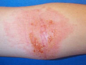
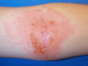
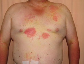
- Acute dermatitis: Evidence of intensely erythematous (reddened) skin, associated with exudation (weeping) and noticeable inflammation.
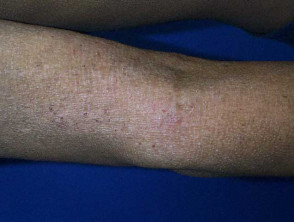
- Subacute dermatitis: Shows a mixed presentation with dry and reddened skin, acting as an intermediate stage between the acute and chronic presentation.
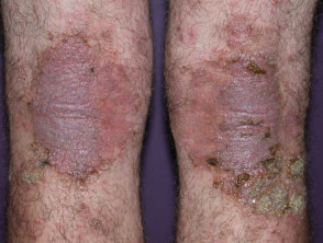
- Chronic dermatitis: The skin tends to darken, dry out excessively, and develop significant thickening, presenting marked furrows due to persistent scratching, a phenomenon known as lichenification.
- Infected dermatitis: This severe complication is characterized by symptoms such as intense localized pain, increased swelling, appearance of pustules and the development of crusts serous or purulent crusts.
Visualization of the Evolution of Cutaneous Dermatitis
Essential Diagnostic Methods for Addressing Persistent Pruritus
To accurately identify the etiology of pruritus, especially when the dermatitis does not present a clear pattern or if constant recurrences exist, it is essential to employ various advanced diagnostic techniques:
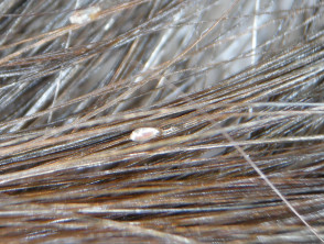
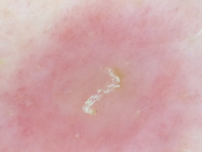
- It is recommended to perform a specific dermoscopic examination of the scalp if it is involved; likewise, if the hands are the affected area, it is crucial to look for signs suggestive of scabies, such as characteristic burrows. burrows.
- It is necessary to take a swab to perform a microbiological examination and obtain a culture Acute bacterial and viral culture, a fundamental procedure when signs of infection such as pustules or the formation of crusts.
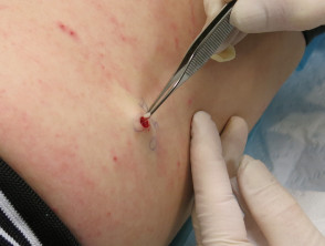
- crusts are observed. In complex cases, a biopsy skin biopsy may be required for detailed histopathology analysis. histopathology. If available, direct immunofluorescence immunofluorescence tests are also used to identify immune deposits.
- When the pruritus is generalized generalized and does not correlate with an identifiable primary skin lesion, the investigation of possible systemic causes is activated. This includes ordering a complete blood count, analysis of iron levels, renal ulceration and hepatic function tests, a thyroid profile, and, occasionally, complementary studies such as a chest X-ray.
An accurate differentiation between the phases of pruritus and dermatitis is essential, as the therapeutic management (which can range from topical emollients to immunosuppressive therapies) depends directly on whether the condition is acute, chronic, or complicated by a secondary infection.
- Chest.
Once the diagnostic process is complete, it is essential to establish an effective management plan. General treatments for these skin conditions usually focus on relieving pruritus and resolving the underlying inflammation.
Therapeutic options for skin pathologies that cause intense itching may include:
- The application of emollients The application of topical lotion and the use of hydrocortisone cream.
- Use of lotions or creams containing 1% menthol to induce a cooling effect on the localized areas affected by pruritus.
- Administration of oral antihistamines to control the reaction.
- The administration of certain tricyclic antidepressants, such as amitriptyline, may be considered in specific cases.
Differential Diagnosis of Intense Skin Itching
To refine the differential diagnosis of intense itching, the following key points should be considered:
- It is crucial to determine whether the itching sensation is generalized over the entire body or restricted to certain specific regions.
- The exact distribution distribution of the pruritus must be accurately assessed.
- It is important to identify whether or not a primary skin rash accompanies the discomfort.
- The presence of and erosions, The presence of erosions secondary excoriation excoriation can significantly complicate the diagnostic process.
Intense Skin Itching Associated with Localized Rash
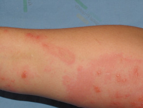
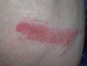
The cutaneous manifestation in this scenario is usually linked to Contact Dermatitis.
- Contact Dermatitis. The final appearance of the lesion is directly determined by the specific substance acting as the causal agent.
- Irritant contact dermatitis Seborrheic is diagnosed more frequently than the allergic form.
- Clinically, an asymmetrical, asymmetrical presentation is typically observed, frequently presenting irregular contours.
- The clinical picture associated with this condition tends to be intermittent in its recurrence.
Contact Dermatitis
Addressing the differential diagnosis of intense itching causes is crucial for implementing the correct treatment and alleviating patient discomfort. A thorough evaluation considering the extent, morphology, and triggering factors will allow for successful dermatological management.
Pediculosis (Head Lice) manifests with:
- Presence of nits (egg capsules) firmly attached near the scalp.
- Observation of tiny blood spots typically located behind the ears.
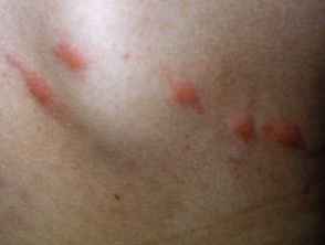
The Insect bites bacterial resistance papular urticaria or resulting papular urticaria present several distinctive characteristics:
- The clinical presentation includes the appearance of wheals, often grouped into elevated papules papules.
- It is common to observe a central central punctum punctum vesicle or the formation of a vesicle in the core of the lesion.
- Lesions tend to be located in areas of skin exposed to the environment, varying according to the type of insect that caused the bite.
Identification of Insect Bites
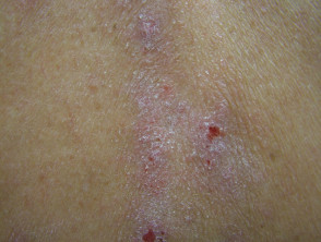
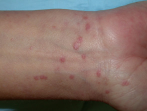
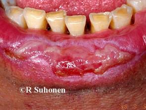
Lichen Planus is a dermatological condition that can manifest anywhere on the body. It is distinguished by the presence of flat, grouped papules that exhibit a polygonal and violaceous (purple) color (forming plaques). Definitive diagnosis of this condition requires confirmation via histopathological biopsy.
Clinical Manifestations of Lichen Planus
An accurate identification of these underlying causes is fundamental to establishing the appropriate treatment and controlling pruritus. If you experience persistent itching or these skin lesions, consult a dermatologist for a specific diagnosis and management plan.
Differential Diagnosis: Exploring Common Pruritic Skin Pathologies
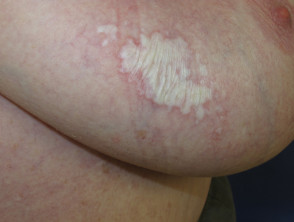
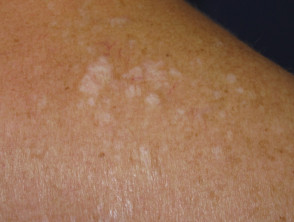
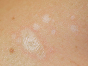
Diagnosis and Manifestations of Lichen Sclerosus
- Although its frequent primary location is in the vulvar and penile region, this condition can manifest in other areas of the body.
- The cutaneous involvement is distinguished by a whitish and dry appearance, characteristic of skin atrophy.
- Over time, it can progress to ecchymosis (appearance of bruises or purpura), blister development, tissue resorption, and subsequent scarring.
- Definitive confirmation of the diagnosis is obtained through histopathological analysis of a skin biopsy.
Key Clinical Images of Lichen Sclerosus
Characteristics of Chronic Simple Lichen (Lichen Simplex)
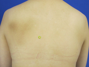
- This presentation is characterized by localized lichenification, which is the thickening of the skin secondary to the repetitive act of scratching.
- Commonly affected areas include the wrists, ankles, neck, scrotum, or vulvar region.
- In cases where the involvement is more extensive, it can develop bilaterally.
Clinical Patterns of Simple Lichen (Neurodermatitis)
Lichen Simplex
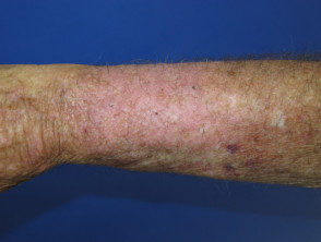
Lichen Simplex
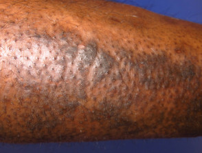
Lichen Simplex
Evaluation of Pompholyx (Dyshidrotic Eczema)
- This condition is characterized by the presence of superficial vesicles vesicles, located mainly on the fingers of both hands and feet, palms, and soles.
Manifestations of Pompholyx (Dyshidrotic Eczema)
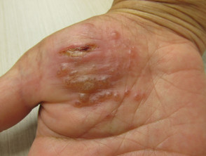
Pompholyx
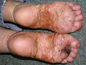
Pompholyx
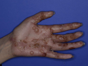
Pompholyx
Considerations on the Venous Eczema
- Typically, this type of eczema starts on one leg at the ankle level, and has the potential to spread to the contralateral leg or generalize through a process of autoeczematization.
- Early signs include…
- Noteworthy manifestation of venous disease: hardening and narrowing of the ankle (lipodermatosclerosis), accompanied by an orange-brown discoloration, a sign of deposited deposition. hemosiderin.
- It often coexists with existing varicose veins.
Venous Eczema and Its Presentations
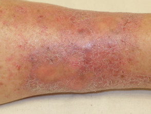
Venous Eczema
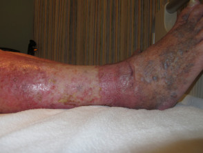
Venous Eczema
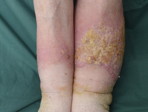
Venous Eczema
Moderate Eczema: Localized Rashes and Itch Intensity
The Discoid, Eczema.
Asteatotic Eczema
- Cracked pavement appearance with red patches.
- It is observed predominantly on the lower extremities (legs).
Representative Images of Venous Eczema
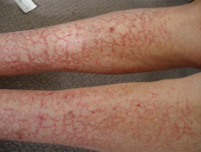
Venous Eczema
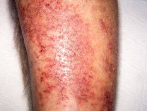
Venous Eczema
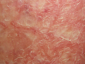
Venous Eczema
Regarding Psoriasis, its distinctive characteristics include:
- The intensity of pruritus associated with psoriasis can be considerable in certain episodes.
- The localized form most frequently affects the scalp, elbows, knees, or the palmoplantar surfaces (palms and soles of the feet).
Visual Characteristics of Psoriasis
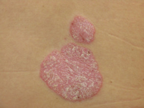
Psoriasis
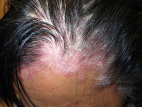
Psoriasis

Psoriasis
Seborrheic Dermatitis: this condition is distinguished by the following locations and appearance:
- Involvement of the scalp with hair, as well as the eyebrows and the hairy areas of the chest.
- It is located in the skin folds, specifically behind the ears, the nasolabial fold, and the axilla.
- It presents a pinkish coloration.
It is essential to complete the analysis of these skin conditions to develop an exhaustive differential diagnosis and apply targeted therapies, understanding that the correct identification of these lesions is the key to effectively managing pruritus and dermatitis.
- Salmon and a characteristic scaly appearance.
Illustrative Images of Seborrheic Dermatitis
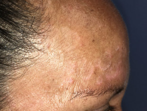
Seborrheic Dermatitis
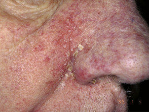
Seborrheic Dermatitis
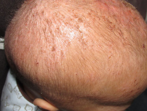
Seborrheic Dermatitis
Evaluation of Intense Pruritus with Generalized Skin Rash
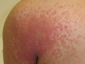
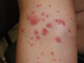
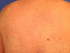
Autoeczematization is considered, which is described below:
- It presents clear signs of associated dermatitis.
The precise identification of the underlying etiology of generalized pruritus and rashes is crucial to establishing the most effective dermatological treatment and providing significant relief to the patient.
- The initial manifestation frequently suggests dermatitis of venous origin.
- Alternatively, it may present as a late consequence of a focal fungal infection, such as tinea pedis (athlete's foot).
Analysis of Autoeczematization and Associated Blistering Lesions
Autoeczematization
Autoeczematization
Autoeczematization
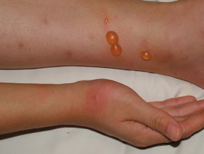
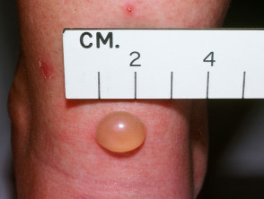
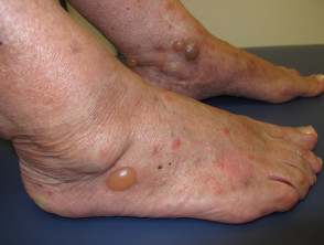
Bullous Pemphigoid
- It primarily affects the elderly, frequently with some degree of cognitive decline.
- Its initial manifestation may resemble eczema or a urticarial reaction.
- It is distinguished by the appearance of large, tense blisters.
- Definitive diagnosis generally requires confirmation via skin biopsy.
Diagnostic Process of Bullous Pemphigoid
Bullous Pemphigoid
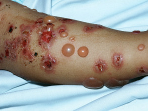
Bullous Pemphigoid
Rigorous diagnostic approach in cases of intense pruritus and generalized rashes, whether due to autoeczematization or underlying conditions like bullous pemphigoid, is fundamental for therapeutic success and improvement in the patient's quality of life.
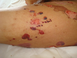
Bullous Pemphigoid
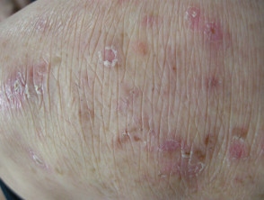
Dermatitis Herpetiformis
- It manifests with itchy vesicles and papules that are rapidly excoriated (scratched).
- Definitive diagnosis requires a confirmatory biopsy.
Characteristics of Dermatitis Herpetiformis
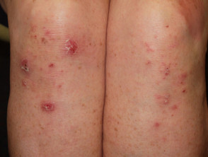
Dermatitis Herpetiformis
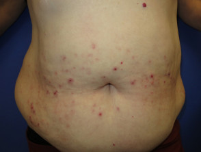
Dermatitis Herpetiformis
Dermatitis Herpetiformis
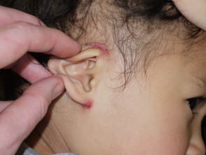
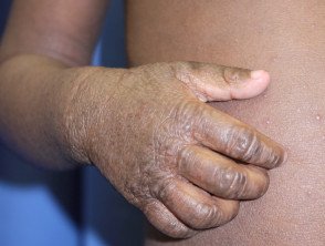
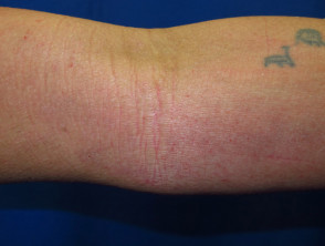
Atopic Dermatitis Atopic
- Primarily located in the flexion, flexural areas symmetrical.
- , presenting a symmetrical pattern. It is frequently associated with notably dry skin.
Atopic Dermatitis (Eczema): Symptoms and Location
Atopic Dermatitis
Atopic Dermatitis
Atopic Dermatitis
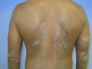
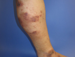
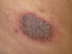
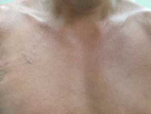
Discoid Eczema
- Typically bilateral presentation, but not necessarily symmetrical.
- It is characterized by the appearance of well- rounded plaques (discoid).
Discoid Eczema: Characteristics and Presentation of Lesions
Discoid Eczema
The correct identification of these pruritic skin manifestations is fundamental to establishing an accurate diagnosis and applying the appropriate dermatological treatment.
Discoid Eczema
Discoid Eczema
Discoid Eczema
Erythroderma
- It involves extensive involvement of the body surface, compromising more than 85% of the skin.
- It can manifest as a severe exacerbation of eczema, psoriasis, or appear as a de novo de novo (new) condition.
- It is essential to consider other important differential diagnoses, such as pityriasis rubra pilaris, certain forms of cutaneous lymphoma lymphoma, or severe drug reactions.
Erythroderma: Urgent Diagnostic Considerations
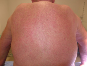
Erythroderma
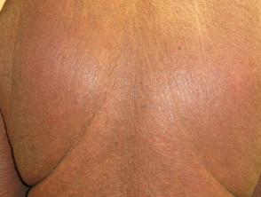
Erythroderma
Lichen Planus
- Involvement of skin and mucous surfaces.
- Presence of violaceous, polygonal, firm plaques grouped on wrists, shins, and lower back.
- White lace pattern visible on the oral mucosa.
- It can manifest as painful erosions on the tongue, vulva, vagina, and penis.
- Diagnostic confirmation is done via biopsy.
Diagnosis and Key Characteristics of Lichen Planus
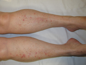
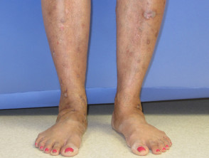
Mycosis Fungoides (T-Cell Lymphoma)Pleomorphic Small/Medium CD4+ Primary Cutaneous T-Cell Lymphoma)
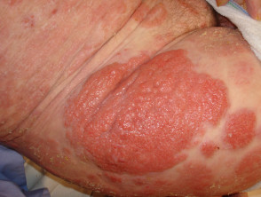
- It gradually evolves from rounded patches, plaques, or, occasionally, nodules, nodules scaly y annular.
- , showing a scaly and annular appearance. It presents diverse clinical morphologies morphologies throughout its progression, hindering early recognition.
- The itching (pruritus) is intense and tends to worsen at night.
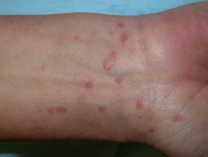
- Distinctive clinical findings include the observable presence of tunnels or burrows, typically located between the fingers, in the wrist folds, or in other areas of thin skin.
- Due to constant scratching, there is a high probability of developing superimposed secondary skin infections.
- The skin rash may manifest as papules in areas such as the armpits, groin, and penis.
- On the trunk, it is common to observe a polymorphous rash (presenting various shapes).
- In cases of immunocompromised patients or the elderly, scabies can evolve into a more severe presentation, known as crusted or scaly scabies.
Key Clinical Characteristics of Mycosis Fungoides
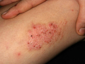
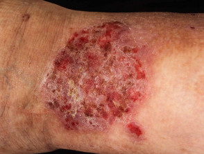
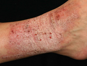
Neurodermatitis, also known as chronic simple lichen, presents specific skin manifestations.
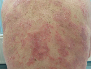
- It is distinguished by the presence of multiple lichenified plaques, a direct result of excessive chronic friction and scratching.
Neurodermatitis: The Phenomenon of Lichenified Skin
Neurodermatitis
Neurodermatitis
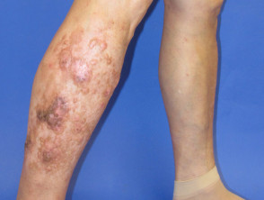
Nodular Prurigo is characterized by a particular skin symptomatology.
- Bilateral nodules appear on the extremities, whose morphology may resemble that of keratoacanthomas.
- This condition also exhibits a predominantly papular presentation variant.
Nodular Prurigo: Clinical Characteristics and Morphology
Prurigo Nodularis
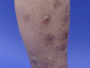
Prurigo Nodularis
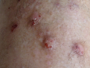
Prurigo Nodularis
Scabies, or Scabiosis, reveals specific clinical signs that aid in its identification.
- It is essential to look for visible tunnels or burrows, which are commonly located between the fingers and in the wrist folds.
- There is a high probability that intense scratching will cause a secondary infection on the affected skin.
- The initial rash includes the appearance of papules in sensitive areas such as the armpits, groin, and penis.
- On the trunk, the presentation is typically a polymorphous rash (with lesions of various shapes).
- In vulnerable groups, such as elderly patients or those with compromised immune systems, scabies can evolve into a severe manifestation characterized by scale and hyperkeratosis (crusted scabies or Norway scabies).
Understanding these detailed clinical manifestations is crucial for differentiating between the various etiologies of chronic pruritus and establishing the appropriate dermatological treatment.
- It presents skin eruptions (hives) scaband often crusting) in areas such as the interdigital space, elbows, and scalp.
Key Differential Diagnosis for Scabies
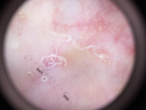
Scabies
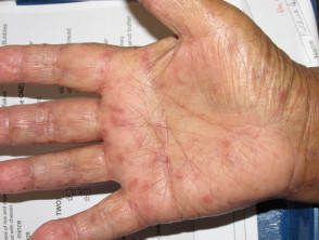
Scabies
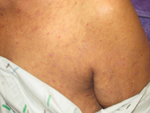
Scabies
The Juvenile Transient transient, commonly known as Grover's Disease, presents a distinctive symptomatology:
- Its appearance is predominantly centered on elderly males.
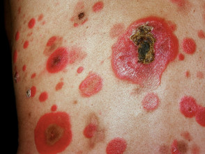
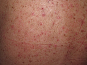
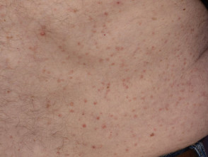
- It manifests through erythematous and crusted papules, located mainly on the mid-trunk.
- Excessive sweating can function as a precipitating or exacerbating factor of the symptoms.
- The distribution of the lesions is usually symmetrical, affecting areas such as the scalp, shoulders, elbows, buttocks, and knees.
Detailed Analysis of Transient Acantholytic Dermatosis
For correct management of Grover's Disease (transient acantholytic dermatosis), it is fundamental to establish a rigorous differential diagnosis, given its specific clinical presentation in the geriatric population. It is important to continuously review morphological presentations to distinguish these complex pruritic conditions.
Transient Acantholytic Dermatosis: Visual Exploration

Diagnostic Criteria for Urticaria:
- Classification by duration: Acute Urticaria (clinical manifestations less than 6 weeks).
- Classification by duration: Chronic Urticaria (persisting for more than 6 weeks).
- Types of Urticaria: Can be spontaneous or inducible, inducible wheals patterns.
- , always defined by the appearance of characteristic wheals. Signs such as blisters, marked dryness (xerosis), or significant scaling are excluded.
- Physical response: Rubbing the skin generates a distinctive linear line or wheal linear wheal.
, indicative of dermographism.
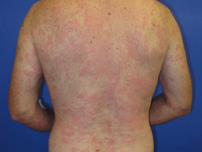
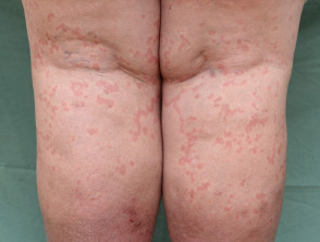
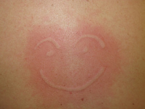
Exploring the Clinical Manifestations of Urticaria
Psoriasis
- Differential Diagnosis: Generalized Pruritus vs. Skin Rashes.
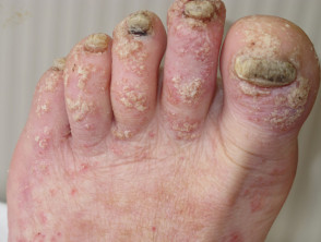
- Psoriasis circumscribed scaly plaques The intensity of pruritus associated with psoriasis is variable and can become considerably intense in some cases. Its characteristic clinical presentation includes well-.
- circumscribed plaques.
that exhibit an erythematous background (reddened) crowned by whitish or silvery scales. The distribution can be focal, limited to specific areas, or extend generally covering large expanses of skin.
Distinctive Characteristics of Cutaneous Psoriasis.
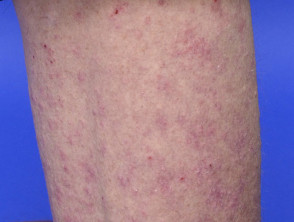
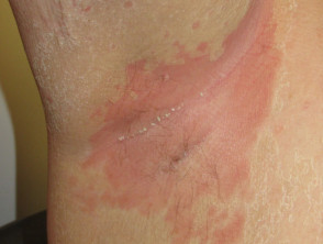
Establishing a clear distinction between predominantly pruritic conditions, such as urticaria, and those that form well-defined plaques, such as psoriasis, is fundamental to achieving an accurate dermatological diagnosis. Precise identification of the elementary lesions and the temporal sequence of symptoms are key to determining the most effective therapeutic approach. eczema
- Xerotic.
Eczema
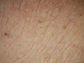
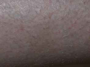
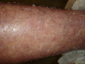
The skin tends to be very dry or dehydrated.
Cutaneous Xerosis: Exploration of Causes and Characteristics neuropathic symptoms. o ) of.
Localized Itching on Skin Without Visible Rash: Neurogenic Causes.
- Pruritus Focal itching can trigger significant secondary lesions, such as erosions from excessive scratching, purpura, chronic simple lichen, or secondary skin infections. Often, the underlying localized pruritus stems from a neuropathic or.
- Meralgia neurogenic origin. outer thigh.
- Notalgia If irritation is specifically concentrated on the scalp, a detailed inspection is essential to look for pediculosis (lice) and their nits (eggs attached to the hair).
- Brachioradial pruritus.
- : Localized involvement in the arm area.
- Meralgia.
: Irritation on the lateral side of the thigh.
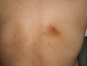
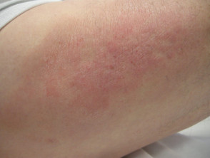
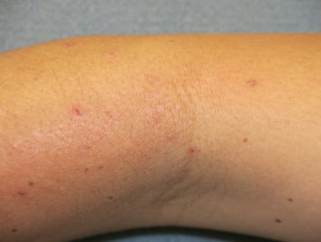
Notalgia paresthetica.
: Pruritus confined to the scapular region (back bone).
Brachioradial Pruritus
Cheiralgia paresthetica
: Manifests on the lateral portion of the hand.
Vulvar pruritus
- : Intense itching in the female genital region.
Scrotal pruritus Systemic : Chronic irritation observed in the male scrotum.
- Understanding Localized Neuropathic Pruritus.
- Cholestasis Determining the precise location and type of pruritus is fundamental to discerning whether the etiology is inflammatory or of nervous origin. This distinction facilitates a more accurate diagnosis and therapeutic approach to mitigate the imperative need for self-mutilation by scratching.
- Iron deficiency.
- Diagnosis of Generalized Pruritus on Skin Without Visible Rash.
- Hyperthyroidism.
- Brachioradial Pruritus.
- Small fiber neuropathy Differential Exploration When Faced with Generalized Pruritus Without Primary Lesions.
- When evaluating generalized itching without an evident primary skin rash, it is imperative to investigate thoroughly, looking for discrete signs such as the possible presence of tiny 'burrows' that could indicate underlying scabies.
- Itching can occur in special circumstances, such as during pregnancy (Pruritus/Prurigo of Pregnancy), where:.
The patient is pregnant.
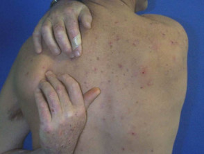
Chronic and persistent pruritus often signals a
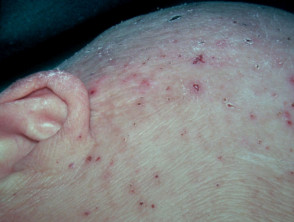
Breast cancer
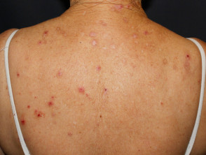
Iron Deficiency
Systemic origin (Term with ID 380). Therefore, investigating a wide range of internal conditions is recommended:.


