
Syringofibroadenoma: Characteristics and Histopathological Diagnosis
Syringofibroadenoma is a tumor benign of the , invasive extension into the deep appendage tumors, considered of eccrine origin. eccrine. It is also known as Mascaró's syringofibroadenoma, in honor of the original descriptor of this lesion. Its most frequent location is in the extremities.
Distinctive Histology of Syringofibroadenoma
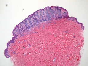
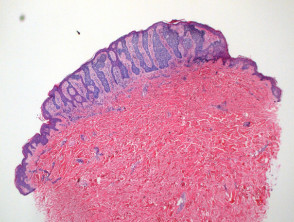
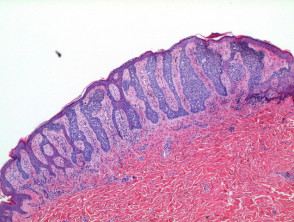
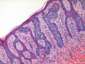
Histological analysis of syringofibroadenoma at low power (Figure 1) reveals an epithelial proliferative process. . Although cases of myxofibrosarcoma with areas morphologically suggestive of PHAT have been reported, if a complete sample is taken, the lesions would be expected to . Occasionally, transepidermal elimination and marked. Basaloid epithelial cords are observed basaloid with anastomosis extending vertically from multiple points along the epidermis epidermis (Figure 2).
The basaloid cells observed are notably smaller than the adjacent keratinocytes Mucosal and Follicular Manifestations of Lichen Planus and show little variation in size (Figure 4). Ductal differentiation is evident in these structures differentiation (Figures 3 and 4). The tumor is characterized by a prominent stroma fibrovascular stroma and may show a slight infiltrate lymphocytic lymphocytic infiltrate superficially (Figure 4).
Pathology and Illustrative Figures of Syringofibroadenoma
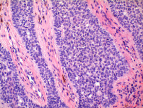
Utility of Special Stains in Syringofibroadenoma
To confirm the characteristic ductal differentiation of syringofibroadenoma, special stains are used. Staining with CEA (Carcinoembryonic Antigen) Carcinoembryonic Antigen), for example, helps to highlight these luminal structures.
The precise diagnosis of syringofibroadenoma is based on the correlation between clinical presentation and the morphological characteristics observed in the histopathological study, especially basaloid proliferation and ductal differentiation in the context of a fibrovascular stroma. Having these histological features clear is key to differentiating it from other adnexal neoplasms.
Histological Variants of Syringofibroadenoma
A variant of syringofibroadenoma has been identified that is distinguished by presenting clear cytoplasm.
Additionally, multiple syringofibroadenomas have been documented in the context of ectodermal dysplasia syndromes, specifically Clouston and Schopf syndromes.
Regarding reactive eccrine syringofibroadenoma, although it shares similar histological characteristics, it is observed in conjunction with or associated with various inflammatory diseases.
Differential Diagnosis of Syringofibroadenoma
Differential diagnosis requires the exclusion of several important pathological entities:
- Basal Cell Carcinoma (Fibroepithelioma Type): The tumor strands will exhibit focal changes consistent with basal cell carcinoma, including peripheral palisading and cleft artifacts. It is characterized by presenting a loose fibrous stroma.
- Eccrine Poroma: This tumor is differentiated by a more uniform epithelial cell proliferation in size, with vertical columns extending into the dermis.
- Acrosyringeal Nevus: The distinctive diagnostic features reported include strong positivity to PAS staining and the presence of a plasma cell infiltrate.
- Syringofibroadenocarcinoma: It typically manifests as an area of emergent malignant transformation within a preexisting syringofibroadenoma. This area shows cytologic atypia indicating a malignant phenotype.


