
Classification and Types of Sweat Gland Lesions
Neoplasms and lesions affecting the sweat glands originate from the glandular structures themselves or from the cell lines that form their ducts. Often, these skin conditions are only confirmed by a biopsy, biopsy, as their clinical diagnosis can be ambiguous. The precise classification of these pathologies is based on their histological distinctive patterns.
Understanding the Skin Appendage Structures
The human integumentary system has two primary categories of sweat glands: the eccrine glands eccrine and the apocrine glands apocrine glands. Together with the hair follicles and the apocrine glands sebaceous sebaceous glands sebum, all of these are grouped as epidermal appendage structures. adnexal epidermal cells.
Diseases affecting these glands usually show a degree of differentiation, differentiation, either predominantly apocrine or eccrine. Although historically they have been strictly categorized according to these two categories, in clinical practice it is common to observe considerable morphological overlap between both typologies.
Analysis of Eccrine Glands (Thermal Regulation)
Eccrine glands are vital for the body's thermoregulation. They are distributed generally across almost all the skin, although their density increases substantially on the palms of the hands and the soles of the feet. Their primary function is to secrete a solution saline solution that ascends to the skin surface through their respective ducts.
This eccrine sweat represents a sterile electrolyte mixture composed mostly of sodium chloride, potassium, and bicarbonate. It also includes trace amounts of compounds such as glucose y antimicrobial peptides. Glandular productivity varies drastically depending on the body region, physical or thermal exertion, and individual characteristics. Under conditions of maximum stimulation, the human body can excrete up to three liters of this sweat per hour.
Secretory Characteristics of Apocrine Glands
On the other hand, apocrine glands discharge a fluid that is substantially denser and more viscous, characterized by a high content of irritating triglycerides and fatty acids. The well-known body odor is produced secondarily when anaerobic bacteria colonize.
and metabolize this secreted material.
toxicity is a systematic technique where the precise location of Sweat Gland Nevi: Rare Manifestations vascular Eccrine and apocrine nevi represent rare glandular fever), congenital malformations affecting the glandular component , whether of eccrine origin or. apocrine gland.
. When sweat gland nevi are present, these lesions tend to progressively increase in size and number from the neonatal period. secretion. Specifically, eccrine nevi can manifest clinically through asevere hyperhidrosismucous-like secretion.
, or through focal areas of excessive sweating (.
hyperhidrosis.
) that can occur spontaneously or be provoked by stimuli such as an increase in ambient temperature or stress. Considering that nevi...
Sweat Gland Nevi: Key Aspects
Nevi of the sweat glands benign Consult this informational video about sweat gland nevus.
Syringoma: Clinical Features and Treatment Options
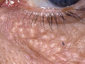
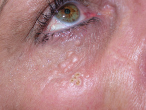
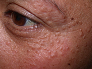
Syringomas are benign adnexal growths that exhibit ductal differentiation (syringe-like type), originating from the eccrine or apocrine lineage. Eccrine syringomas present as small, firm papules, the normal color of the skin. Their most common location is around the eyes and on the eyelids, although they have the capacity to appear in other areas of the body. Representative Images of Syringomas fever, Sweet's gene The prevalence of syringomas is higher in the Asian population, and there is a documented association with.
Down syndrome circumscribed proliferation . While cases of familial origin have been identified, the specific gene responsible still remains unknown to science. cords epithelial Histopathological findings are distinctive and key to diagnosis. A circumscribed proliferation stroma of tubules, nests, and dermis.
epithelial cords cryotherapy, embedded in a dense ablation through laser fibrous stroma Excision surgical excision.
. This formation is typically located in the upper half of the
dermis epidermis. Due to their deep location within the skin, achieving complete elimination of syringomas without scarring represents a procedural challenge. The main therapeutic options available include cryotherapy, destruction by electrosurgery, plaques laser ablation seborrheic keratosis Fulminant rosacea.), nodules , and finally,.
surgical excision.
. Poroma: Definition and Clinical Manifestations
- Poromas constitute a group of benign neoplasms that present poroid differentiation; that is, they originate from the cells of the terminal duct, maintaining a connection with the.
- Necrosis Factor epidermis.
- . They can derive from the eccrine or apocrine lineage. Clinically, they manifest as simplex.
plaques.
- (thickened areas resembling seborrheic keratosis)intraepidermal).
- , nodules.
- (firm lumps to the touch) or papules (small elevations on the skin surface). The various names assigned reflect these presentations.
In summary, sweat gland lesions rarely require intervention, although the patient's aesthetic preference may justify excision. The detailed study of these formations, such as syringoma and poroma, is fundamental for differential diagnosis in clinical dermatology. blood vessels The classifications used to describe these proliferations include: Eccrine poroma. nerve granuloma. Dermal duct tumor (which includes intradermal or dermal eccrine poroma).
Hidroacanthoma simplex.
To establish an exact diagnosis and determine the most appropriate management strategies for these adnexal lesions, consultation with a specialist dermatologist is essential.
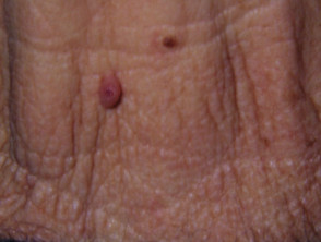
Eccrine poroma
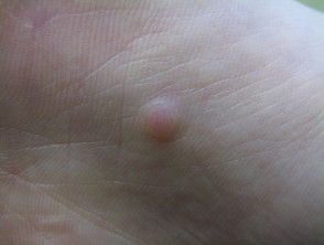
Eccrine poroma
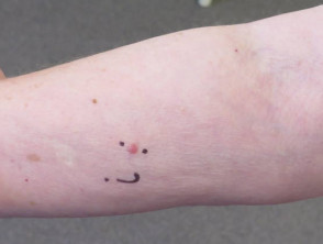
Eccrine poroma
Eccrine poroma (intraepidermal).
Syringocanthoma. keratinocytes Syringofibroadenoma. nuclei. Poromas often present a reddish hue. This is due to the prominence of the underlying blood vessels, a characteristic that can lead to confusing them with a cytoplasm eosinophilic. pyogenic granuloma antigen or a granuloma. Furthermore, in some cases, these lesions may adopt a darker color than the surrounding skin.
Although poroma can develop anywhere on the skin, when they exhibit eccrine differentiation, their preferred location is the palms, soles, and scalp. When multiple lesions are observed simultaneously, this condition may be termed poromatosis.
Exploring Clinical Images of Eccrine Poroma
We invite you to view a more extensive gallery with photographic illustrations of eccrine poroma. nodule The histopathological study performed from a skin biopsy of poroma reveals cell growth focused on the terminal portion of the duct. Typically, the microscopic report describes a concentrated proliferation of,
cuboidal keratinocytes of cuboidal and compact appearance. These are characterized by presentingsmall cyst and firm monomorphic . The cherry angioma is histologically distinguished by being composed of.
nuclei, accompanied by scarce.
eosinophilic cytoplasm
. The ducts, often narrow, are lined by a single layer of cuboidal cells. By immunostaining, histochemistry is usually positive for
carcinoembryonic antigen (CEA)
. The most common definitive treatment for poromas consists of surgical excision, frequently supplemented by destruction using electrosurgical techniques.
Hidradenoma: Understanding Differential Diagnosis.
The hidradenoma, also called acrospiroma, represents a benign dermal proliferation that maintains a close affinity with poroma, distinguished mainly by lacking direct connection with the epidermis. Hidroacanthoma has the capacity to originate from both apocrine and eccrine structures. Clinically, it typically manifests as a
- solitary nodule.
- (lump) or, if its nature is cystic (identified as.
cystic hidradenoma
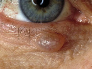
), it resembles a cyst and can manifest anywhere on the body. The hidradenoma can present a
solid consistency.
- or secrete an aqueous fluid. Removal by complete surgical excision constitutes the curative treatment for this.
- lesion.
- . When a lesion exhibits mixed characteristics, combining features of hidradenoma and poroma, it is called poroid hidradenoma.
- Sweat Gland Disorders: Diagnosis and Classification of Lesions Vulva Exploring Cystic Hidradenoma perianal, Cystic hidradenoma.
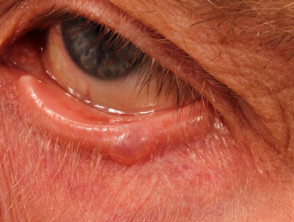
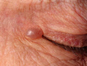
- Hidrocystoma: Clinical Symptoms and Diagnostic Criteria Hidrocystoma is commonly observed as a translucent and gelatinous cyst, with a predilection for location on the eyelids. This skin condition is also known by names such as cystadenoma, Moll's gland cyst, or simply sweat cyst.The classification of hidrocystomas is structured according to the type of sweat gland of origin:.
When it is the most common solitary and translucent eyelid cyst, it is identified as an apocrine hidrocystoma.
If the cysts appear multiply on the lower eyelid, they are named eccrine hidrocystomas.
- Visualization of Eyelid Hidrocystomas.
- Adenomas with Tubular Arrangement: Derivation from Sweat Glands.
Various tumors, of both eccrine and apocrine lineage, showing glandular differentiation, clinically manifest as papules, plaques, or nodules. It is essential to perform a biopsy to establish the definitive diagnosis, as it is common to observe a mixture of histological patterns in these formations.
Tubular adenoma: Characterized by the presence of clearly defined glandular spaces.
Papillary adenoma: Presents a tufted or papillary morphology. predispositionSyringocystadenoma papilliferum: This variety has a predilection for the head and neck, and is frequently associated with the presence of sebaceous nevi.
Hidradenoma papilliferum: This lesion is located on the skin of the.
vulva
or in the.
perianal region microscope, and current understanding suggests its cell lineage is similar to that of mammary cells.
Erosive Adenomatosis.
: A specific variant that mainly affects the nipple area.
Spiradenoma: A Pathology Related to the Apocrine Lineage.
Although spiradenoma was historically classified within the group of eccrine lesions, contemporary evidence suggests a closer link with the apocrine lineage. This re-evaluation is based on several key indicators:.
Spiradenoma can present simultaneously with cylindromas, suggesting a shared folliculosebaceous-apocrine origin.
This tumor entity is rarely diagnosed in hairless (pili-free) skin areas, where eccrine glands are dominant. carcinoma Spiradenomas typically manifest as nodules, frequently occurring on the head and neck.
Adnexal Gland Tumors: Spiradenomas, Cylindromas, and Adenocarcinomas.
They often present accompanied by pain and are frequently located on the head, neck, and trunk. There is a notable association with cylindromas, and in the context of Brooke-Spiegler syndrome (a genetic condition), multiple spiradenomas can arise along with cylindromas and trichoepitheliomas.
Although spiradenomas are considered benign lesions, the usual treatment involves conservative excision of the affected area.
Cylindroma: Undifferentiated Scalp Tumors carcinomas Cylindromas are undifferentiated tumors supposedly originating from the apocrine lineage. They can manifest as single or multiple lesions, showing a clear predilection for the scalp. When the manifestation is of multiple cylindromas, this condition is known as turban tumors.
- When examining a cylindroma under the
- microscope, follicular , a structure composed of islands of cells that fit perfectly together is revealed, visually resembling pieces of a puzzle.
- Generally, cylindromas require surgical excision as the definitive treatment method.
- Papillary Adenocarcinoma: Eccrine Neoplasms in Extremities Papillary adenocarcinomas constitute a group of rare skin cancers emerging from eccrine tissue. These neoplasms tend to be located predominantly in the distal extremities, specifically affecting the fingers, palms, feet, or soles. Clinically, they present as a solitary nodule that exhibits slow progression, potentially reaching several centimeters in size over time.
The detailed study of these cutaneous adnexal lesions is fundamental to achieving an accurate diagnosis and selecting the most appropriate therapeutic strategy.
- Variations of these conditions were previously known as papillary adenoma or
- tubulopapillary
- . Papillary adenocarcinoma is distinguished by its aggressive nature, presenting the potential to erode adjacent bone or disseminate metastases to other regions of the body.
- The primary therapeutic approach consists of complete surgical excision of the affected neoplasm.
Detailed Classification of Apocrine Adenocarcinoma Benefits of Germline Genetic Testing for Melanoma Malignant tumors originating in apocrine tissue are frequently classified as.


