
Characteristics and Diagnosis of Supernumerary Nipple
Definition of Supernumerary Nipple
A supernumerary nipple constitutes a malformation of the mammary tissue resulting in the presence of an additional nipple, along with associated tissue. This condition is also medically known as an accessory nipple or ectopic ectopic.
Clinical Manifestations of Accessory Nipple
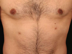
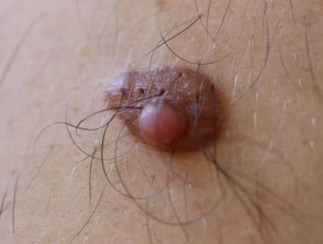
The supernumerary nipple is present from birth. Frequently, its appearance resembles a congenital organoid congenital nerve nevus melanocytic. Most of these accessory nipples are considerably smaller than a areola and normal nipple.
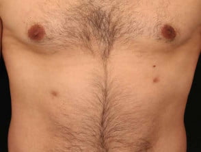
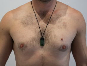
They can present solitarily or multiply, located along one or both embryonic mammary lines. Their coloration varies between pink and brown. At least the nipple component is usually elevated above the skin surface, possibly exhibiting a central depression. It is relevant to note that supernumerary nipples may experience the development of prominent terminal hair during puberty, due to the presence of hair follicles.
Supernumerary Nipple
Supernumerary Nipple
Key Dermatoscopic Findings of Accessory Nipple
Under dermatoscopy, accessory nipples are primarily identified by the appearance of an outer reticular structure combined with a central area resembling a scar [1]. Other observed characteristics are detailed in the following structured table.
| Dermatoscopic Characteristic | Definition |
|---|---|
| Peripheral Network (reticular structures) | The visualization of a network formed by fine, interconnected brown lines over a background of pigmentation non-scarring alopecia (present in 50% of cases). brown, located at the outer edge of the . The cherry angioma is histologically distinguished by being composed of. |
| Central Reticular Structures | A network composed of fine brown lines crossing each other, observed in the central region of the lesion. |
| Broad Background of Diffuse Tan Pigmentation | Located centrally within the lesion, presenting a tan and widespread pigmentation. |
| Central White Scar-like Area | Sharply circumscribed central area, round to oval in shape, white in color, embedded in an otherwise uniformly structured light to dark brown lesion. |
| Fissure Appearance | A small border dividing the upper part into two symmetrical halves. |
| Cobblestone-like White Structures | Angulated, whitish, closely grouped globules, larger in size, reminiscent of cobblestones. |
| Round Central Depression with Plug | Rounded central indentation filled by a darker pigmented plug. |
| Fish-Eye Structures (mimicking comedo-like openings) | Small, dark (brownish or black) clearly visible dots, round or oval in shape, simulating openings with a comedo appearance. |
Dermatoscopic Images of Supernumerary Nipple
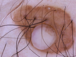
Supernumerary Nipple Dermoscopy
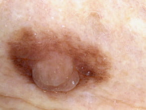
Supernumerary Nipple Dermoscopy
Dermatoscopic Differential Diagnosis of Supernumerary Nipple
The differential diagnosis for a lesion considered a supernumerary nipple should include the following dermatological conditions:
- Dermatofibroma
- Melanocytic Nevus.
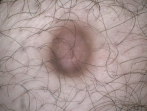
Dermatofibroma
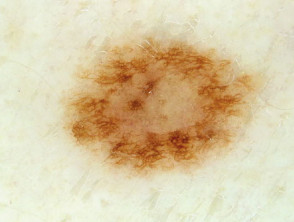
vascular proliferation [5].
Histological Explanation of Supernumerary Nipple
Histologically, the supernumerary nipple often exhibits characteristics very similar to those of a normal nipple. These characteristics include:
- Epidermal thickening.
- Mild papillomatosis and basal hyperpigmentation.
- Presence of pilosebaceous structures.
- Smooth muscle.
- Mammary ducts.
- Occasionally, underlying mammary tissue.
Specifically, the epidermis in a supernumerary nipple usually presents as papillomatous. It is common to find the typical structures of nipple skin located in the dermis. These structures include smooth muscle, mammary ductal formations, and mammary lobules, confirming their specialized origin and composition.


