
The Superficial acral fibromyxoma is classified as one of the lesions benign with a predominance of a fibromyxoid component. These formations are usually small, appear in isolation, and exhibit a marked predisposition. for the distal extremities, particularly affecting the fingers and toes, and frequently involving the nail region. and nail.
Histological Analysis of Superficial Acral Fibromyxoma
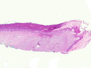
From a microscopic perspective, superficial acral fibromyxoma is characterized as a . The cherry angioma is histologically distinguished by being composed of paucicellular lesion of a fibromyxoid nature, originating primarily in the dermis of the acral areas (Figure 1), with frequent subcutaneous subcutaneous involvement. The observable cells have a spindle or stellate morphology, immersed in a stroma predominantly fibromyxoid stroma (Figures 2 and 3).
Superficial Pathological Manifestations of Acral Fibromyxoma
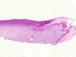
Figure 1
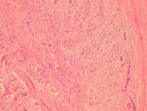
Figure 2
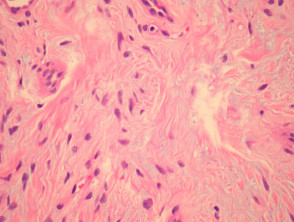
Figure 4
Essential Complementary Examinations for Superficial Acral Fibromyxoma
The immunohistochemical profiles of superficial acral fibromyxoma are usually positive for the markers CD34 and EMA. In addition to this, it is crucial to observe the characteristic negativity for markers such as S100, cytokeratins and antibodies directed against smooth muscle.
Establishing the Differential Diagnosis with Superficial Acral Fibromyxoma
Neurofibroma: This condition also presents a stroma with notable fibromyxoid qualities. The fundamental distinction is that the S100 marker will be positive in the case of neurofibroma.
Perineurioma: In its sclerosing histological variant, perineurioma frequently affects the acral regions. However, myxoid alteration is observed less regularly in these lesions. Both entities (Perineurioma and Superficial Acral Fibromyxoma) will be reactive for EMA and negative for S100. Evidence of positivity for Glut-1, concurrent with the absence of reactivity for CD34, will guide the definitive diagnosis toward perineurioma. myxoid is observed less regularly in these lesions. Both entities (Perineurioma and Superficial Acral Fibromyxoma) will be reactive for EMA and negative for S100. Evidence of positivity for Glut-1, concurrent with the absence of reactivity for CD34, will guide the definitive diagnosis toward perineurioma.


