
Subcutaneous Fat Necrosis of the Newborn: Causes, Symptoms, and Treatment
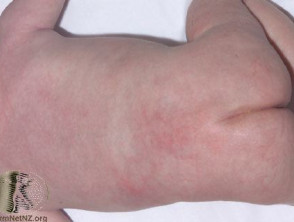
Subcutaneous fat necrosis of the newborn is a rare dermatological condition characterized by the appearance of nodules (lumps) firm, inflamed, and purple nodules in the fatty tissue. This condition generally manifests during the first few weeks after birth and is classified as a form of inflammation of the subcutaneous fat tissue)..
These nodules may present as distinct bumps located on the back, buttocks, and extremities, or they may expand to form large hardened areas. It is important to note that it is considered less severe than neonatal sclerema, another condition that also causes skin hardening in newborn babies.
Images of Subcutaneous Fat Necrosis of the Newborn
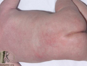
Neonatal Subcutaneous Fat Necrosis
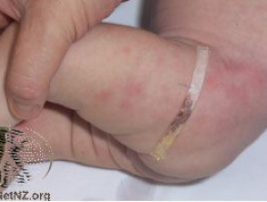
Neonatal Subcutaneous Fat Necrosis
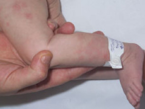
Neonatal Subcutaneous Fat Necrosis
What are the probable causes of this condition?
The exact etiology of subcutaneous fat necrosis is still unknown. However, medical literature suggests that certain intrapartum stressors may increase the probability of its development:
- Fetal stress during the birthing process.
- Conditions of low oxygenation (hypoxia).
- Exposure to cold temperatures.
- History of Cesarean section.
- High birth weight (macrosomia).
- Presence of secondary Presence of concurrent infection.
Despite the presence of these risk factors, it is crucial to understand that most neonates exposed to these circumstances do not develop subcutaneous fat necrosis.
What are the associated complications?
The nodules caused by subcutaneous fat necrosis can cause discomfort or pain. Generally, these remain visible for a period ranging from weeks to months before resolving. Occasionally, resolution may leave a scar of depression or irregular appearance in the affected fatty area.
The most significant systemic complication associated with this condition is hypercalcemia (elevated calcium levels in the blood). Since this is only detected through biochemical analysis, it is essential that neonates diagnosed with subcutaneous fat necrosis be monitored with periodic checks of serum calcium during the first months of life. High calcium levels can manifest as irritability, constipation, difficulty gaining weight, and, very rarely, alterations in heart rate.
Additionally, cases have been documented where episodes of thrombocytopenia (low platelet count) platelet) and hyperlipidemia (elevated lipid concentrations in the serum) are present.
How is the treatment for subcutaneous fat necrosis approached?
The spectrum of treatment for subcutaneous fat necrosis primarily focuses on the active management of hypercalcemia, if it develops. The approach to correcting hypercalcemia may include administering increased fluid intake, using low-calcium diets (restricted dairy), diuretics such as furosemide, the administration of corticosteroids and, if necessary, bisphosphonate therapy.
Fortunately, the inflammation underlying inflammation of the surrounding fat usually resolves spontaneously without the need for specific therapeutic interventions. It is essential to maintain close clinical supervision until the complete resolution of symptoms.


