Mastering Subcision: A Complete Guide to Treating Depressed Scars and Wrinkles
Subcision is established as a highly technical minor surgical procedure, primarily conceived to correct depressed skin scars and persistent facial wrinkles. Formally, this intervention is called subcutaneous incisional surgery.
The method consists of the meticulous introduction of a specialized hypodermic needle through a superficial microincision into the dermis. Using the beveled edge of this instrument, the specialist cuts and separates in a controlled manner the fibrous bands that anchor the scar deeply to the underlying tissue.
The subsequent release of these fibrotic adhesions, combined with neocollagenesis induced by the healing response of the injured tissue, generates a substantial cosmetic improvement in the appearance of the lesion. Subcision is a safe procedure usually performed on an outpatient basis and is generally well tolerated by the patient.
Fundamental Clinical Indications for Dermatological Subcision
The decision to perform subcision must be based on a thorough clinical evaluation, considering the specific type, location, severity of the scars, and the realistic goals of both the patient and the medical professional.
The subcision technique offers versatility to address various dermal conditions, including:
- Depressed scars that exhibit mobility (sequelae of acne, surgical trauma, or previous injuries).
- Indented scars resulting from acne (excluding deep atrophic varieties such as «ice pick» scars), chickenpox, or minor trauma.
- Depressions observed in skin graft areas.
- Deep facial wrinkles considered as furrows.
- Chronic panniculopathy (cellulite) that causes localized dimpling or indentations.
In-Depth Analysis of Post-Acne Atrophic Scars
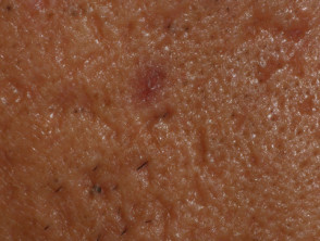
Rolling scars
Absolute and Relative Contraindications for Subcision Application
Despite being a minimally invasive procedure, there are specific clinical conditions that determine whether subcision is the safe and appropriate treatment for the patient:
- Documented history of keloids or excessive hypertrophic scarring.
- Current or recent use (within the last 12 months) of systemic retinoid therapies (such as isotretinoin or acitretin).
- Existence of active coagulation disorders or coagulopathies that prevent proper hemostasis.
- Presence of active infections, both bacterial and viral, in the skin region designated for treatment.
Advanced Classification of Acne Scars: Towards Focused Treatment
To achieve objective evaluation and quantify post-treatment improvement, the severity of acne scars is frequently classified using standardized tools, such as the qualitative system developed by Goodman and Baron.
1. Macular Scars
These are flat areas of color alteration, with no changes in the texture of the dermal relief. They are distinguished by being erythematous, hyperpigmented, or depigmented.
Scar Classification and Subcision Applicability
Macular scars are characterized as flat marks that clinically manifest in three presentations: erythematous (reddish), hyperpigmented (brown due to excess melanin), or hypopigmented (lighter or white areas). It is essential to understand that the subcision procedure is not primarily focused on treating this specific type of superficial flat lesion.
These marks do not represent an alteration of the tissue contour, a situation presented by other degrees of scarring; their main characteristic lies in the modification of skin color.
Scar Classification: Mild to Severe Grades
Mild Atrophic or Hypertrophic Scars
Scars classified as mild atrophic (indented or thin) or slight hypertrophic (thick) usually go unnoticed at a social distance of 50 cm. Generally, these imperfections can be effectively concealed with makeup or by the subtle shadow generated by shaving the beard in men, or by natural body hair in non-facial areas.
Moderate Atrophic or Hypertrophic Scars
When atrophic or hypertrophic scars reach a moderate grade, they become perceptible at social distances greater than 50 cm. In these cases, makeup or hair shadow (shaved beard or extrafacial body hair) is not sufficient to cover them adequately. However, regarding atrophic scars, it is still feasible to achieve some degree of superficial leveling by manually stretching the skin.
Severe Atrophic or Hypertrophic Scars
Atrophic or hypertrophic scars considered severe are clearly visible even at wide social distances, exceeding 50 cm. These lesions are not easily hidden with cosmetic products or the normal shadow of hair, nor is it possible to flatten their surface through manual manipulation or tension of the skin.
Technical Description of the Subcision Procedure
Below are the fundamental technical steps that make up the subcision procedure, used for the effective treatment of specific atrophic scars. This treatment seeks to release underlying fibrous adhesions.
- The area to be treated undergoes an exhaustive cleaning to eliminate any residue of makeup or dirt accumulated on the skin.
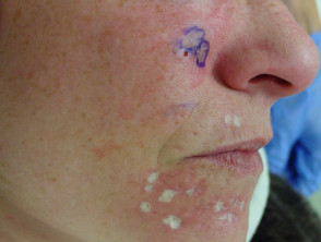
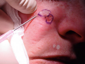
- To ensure precise demarcation of the scar depressions, it may be necessary to outline their borders using a surgical marker and adjusting the ambient lighting (e.g., overhead ceiling light).
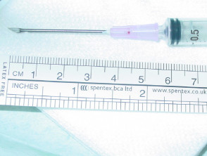
- The necessary local anesthetic is infiltrated, completely covering the area designated for treatment.
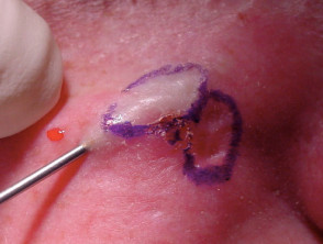
- A three-beveled needle (usually 18 or 20 gauge), or alternatively a Nokor needle (Fig. 3), is introduced, maintaining an acute angle and in an to the sebaceoma (Figure 6). Figure 7, on the other hand, shows less maturity in the follicular structures and the presence of dilated to the scar. The bevel must maintain an upward orientation and remain parallel to the skin surface. For treating superficial wrinkles or smaller scars, the use of finer needles (25-27 gauge) is recommended.
- The needle is introduced through the dermal plane dermis and actively manipulated using a retrogressive and expansive movement, configured like a fan. The success of the release is confirmed by perceiving a characteristic audible snap, indicating the successful sectioning of the fibrous bands in the deep planes of the dermis and the dermal subcutaneous tissue.
- The needle is rotated 90 degrees and the fan-like mobilization is repeated through the affected tissue within the dermis, thus completing the fan subcision action.
- After needle extraction, pressure is applied circumferentially around the exit point to significantly mitigate the appearance of large hematomas resulting from internal bleeding.
- Finally, sustained manual compression is exerted directly over the treated area for several minutes to control the vascular response.
* It is essential to avoid performing the fan movement at excessive depth, inadvertently penetrating below the dermal plane, which is considered a common technical error.
Visualization of the Fan Subcision Technique for Acne Scars
The effective treatment of atrophic scars, especially those moderate or severe presenting significant fibrous anchoring, depends on the precision of the subcision procedure. By releasing these bands, the skin is allowed to elevate upward, notably improving the superficial texture of the affected area.
Subcision is a minimally invasive, highly effective surgical technique designed to release the fibrous anchors that pull the dermal tissue downward, causing depressions characteristic of acne scars. This process notably improves the overall contour of the treated skin.
Post-Subcision Immediate Care and Recovery Protocol
After completing the subcision procedure, it is essential to follow an immediate recovery protocol to ensure proper healing and minimize unwanted side effects:
- Localized pressure and cold therapy (ice) are applied directly to the treated area. The **main objective** is to achieve hemostasis and mitigate the risk of developing extensive bruising.
- Patients have the option to use corrective or cosmetic makeup to conceal the erythema (redness) and ecchymoses (bruises) that are expected sequelae.
- Some clinical professionals recommend the prophylactic administration of antibiotics and drugs Insect stings, such as bees or wasps. to prevent or reduce the severity of potential complications.
It is prudent clinical practice to limit the extent and aggressiveness of manipulations during the initial session. This allows observation of each patient's particular response to the technique, especially relevant in anatomical sites known for having a predisposition predisposition. to generate a hypertrophic or keloid scar response after treatment.
Determining the Number of Sessions Required for Scar Correction
The individual response to subcision treatment varies significantly, strongly influenced by the patient's biological capacity to regenerate new collagen. The precise number of subcision sessions required to achieve the attenuation of a specific depression depends directly on its morphological type, location, how severe the atrophy is, and the intensity applied in each intervention.
Generally, in clinical scenarios where moderate acne scarring predominates, a protocol of three to six visits is considered adequate. It is common to establish a minimum interval of four weeks between each procedure to allow for optimal reorganization of the subdermal tissue.
Potential Risks and Complications Associated with the Subcision Technique
As with any procedure involving manipulation of subcutaneous tissue, subcision presents potential risks and complications that must be discussed thoroughly with the patient before proceeding. These include:
- Appearance of significant bruising due to internal bleeding (although a discreet bruise is considered a normal reaction).
- Experiencing temporary pain or hypersensitivity in the areas subjected to needle insertion.
- Risk, although low, of developing hypertrophic scars (occurring in approximately 5 to 10% of cases) or keloid scars. These adverse responses are more frequent in areas of thin skin such as the periorbital area, the glabella area, the lip border, and the upper lip. periorbital area, the glabella, glabela area lip border and the upper lip.
- Potential for infection, which can often present as a localized episodes of angioedema without hives may originate from angiotensin-converting enzyme (ACE) inhibitors. acne papule papule or a pustule.
- The formation of a pustule.
- The temporary emergence of hyperpigmentation post-inflammatory, post-inflammatory hyperpigmentation, making it essential to recommend rigorous sun protection and avoid direct sun exposure.
- Achieving an insufficient therapeutic response or, directly, observing no improvement in the appearance of the scars.
- There is a risk of injuring deep structures such as a optic nerve blood vessel. or a, temporal blood vessel. This danger increases the closer one works to the bony structures in the mandibular, temporal, and preauricular areas.
Optimized Combination Treatment Strategies with Subcision
The subcision technique proves extremely valuable when adequately integrated into a multimodal treatment plan designed to correct atrophic acne scars. It can be effectively combined with various therapeutic modalities, including:
- Topical retinoid Topical topical (topical application) systems.
- Minimally invasive procedures such as Microneedling (using devices like Dermaroller or Dermapen), which can be safely performed 24 hours after the initial subcision session.
- Chemical peels based on acids, such as 15% Trichloroacetic Acid (TCA), generally recommended every four weeks.
- Application of Trichloroacetic Acid (TCA) at higher concentrations (known as TCA cross).
- Fractionated laser-based laser therapies.
Prognosis and Final Aesthetic Outcomes After Subcision
When subcision is applied to suitable candidates and post-procedure instructions are strictly followed, the achievable aesthetic results are frequently considered excellent. However, it is crucial that the patient understands that total correction of the sequelae may require reinforcement sessions or the repetition of complementary treatments to achieve the desired level of cosmetic improvement. Subcision is a fundamental pillar in the revision of deep acne scars.