Dermoscopy of Squamous Cell Carcinoma

scc1__protectwyjqcm90zwn0il0_focusfillwzi5ncwymjisingildfd-3256100-3207564-jpg-1523679

Table of Contents

¿Qué es el Carcinoma de Células Escamosas Cutáneo?

Cutaneous Squamous Cell Carcinoma, frequently abbreviated as SCC, is a prominent type of cancer non-melanoma skin cancer, specifically of keratinocytic. origin. This condition typically manifests in areas of the skin that have had significant sun exposure. It is crucial to recognize that SCC can become invasive invasive and have the capacity to metastasize Benefits of Germline Genetic Testing for Melanoma, although it is also called squamous cell carcinoma of the skin.

Clinical Manifestations of Squamous Cell Carcinoma

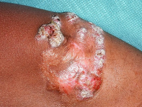
Clinically, cutaneous squamous cell carcinoma usually presents as an nodule elevated, irregular, and keratinous nodule, or as an plaque lesions... (reddish) and firm plaque that often develops ulcers over time.

In many cases, these cancerous lesions emerge from pre-existing skin conditions, such as seborrheic keratosis actinic or invasive carcinoma intraepidermal.

  • The growth of these lesions can extend over weeks or months.
  • Frequently, they develop ulcerated areas.
  • They are often tender or, in some cases, frankly painful to the touch.
  • Their preferred location is sites with high sun exposure, including the face, lips, ears, hands, forearms, and calves.
  • The diameter of the lesions can vary substantially, from a few millimeters to several centimeters.
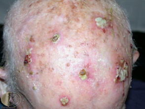
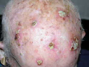
Multiple squamous cell carcinomas visible on a patient's scalp.

Multiple squamous cell carcinomas on the scalp
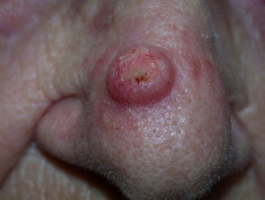
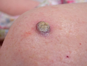
Clinical example of squamous cell carcinoma located on the nose.

Squamous cell carcinoma of the nose

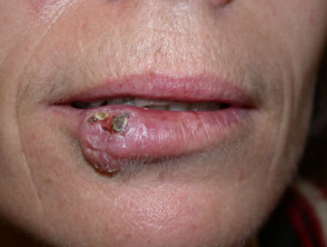
Specimen of squamous cell carcinoma developed on the lip area.

Squamous cell carcinoma of the lip

Representation of a cutaneous squamous cell carcinoma classified as high-risk.

High-risk cutaneous squamous cell carcinoma
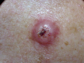
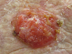
Detailed macroscopic image of a squamous cell carcinoma.

Squamous cell carcinoma

Another visual example of cutaneous squamous cell carcinoma on the skin.

Squamous cell carcinoma

Given its potentially aggressive nature and high frequency in sun-exposed populations, early detection of cutaneous squamous cell carcinoma is fundamental for successful treatment and minimizing the risk of invasive complications.
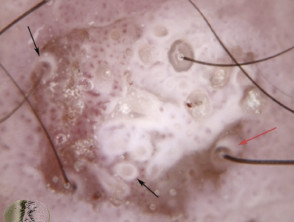
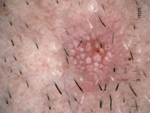
Key Dermatoscopic Features of Squamous Cell Carcinoma

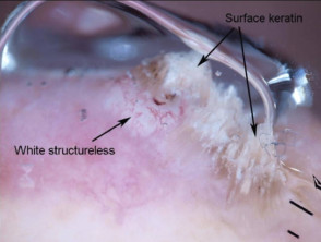
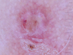
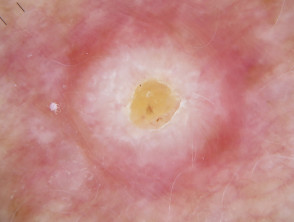
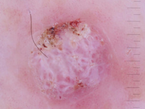
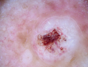
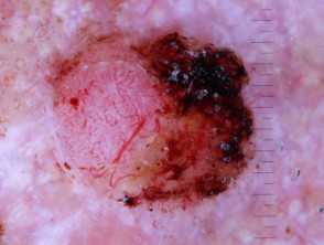
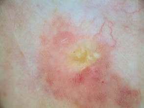
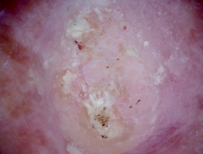
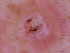
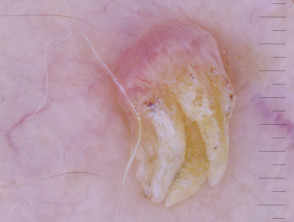
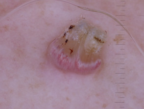
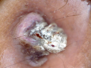
Dermoscopic diagnosis of squamous cell carcinoma (SCC) reveals several distinctive features that help specialists identify this cutaneous neoplasm. These visual manifestations are crucial for differentiating SCC from other lesions.

Dermoscopic features commonly observed in squamous cell carcinoma include:

  • Presence of white circles.
  • Distribution of white areas without an apparent structure.
  • Visualization of loop-shaped blood vessels (anangiectatic vessels).
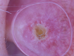
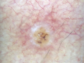
  • Identification of a central keratinous border.
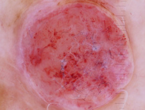
  • A pink or red background, especially prominent in tumors that are poorly differentiated or exhibit rapid growth.
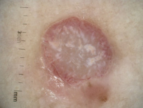
Manifestation of structureless white areas and white circles in dermoscopy of squamous cell carcinoma.

Structureless white areas and white circles in dermoscopy of squamous cell carcinoma

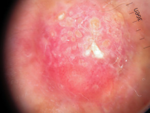
Observation of characteristic white circles during the dermoscopic examination of a squamous cell carcinoma.

White circles in dermoscopy of squamous cell carcinoma

Detail of white circles and the presence of superficial keratin visible via dermoscopy in SCC.

White circles and superficial keratin in dermoscopy of squamous cell carcinoma

Identification of white structures and accumulation of superficial keratin in the dermoscopic study.

White structures and superficial keratin in dermoscopy of squamous cell carcinoma

Visualization of typical loop vessels during the dermoscopic examination of an SCC.

Loop vessels observed in dermoscopy of squamous cell carcinoma

Findings of structureless white areas along with well-defined keratin in the center of the lesion.

Structureless white areas and central keratin in dermoscopy of squamous cell carcinoma

Structureless white areas and superficial keratin in dermoscopy of squamous cell carcinoma

Structureless white areas, central keratin, and loop vessels in dermoscopy of squamous cell carcinoma
Dermoscopy of poorly differentiated squamous cell carcinoma

Dermoscopic Differential Diagnosis of Squamous Cell Carcinoma

The differential diagnosis performed via dermoscopy of cutaneous squamous cell carcinoma (SCC) encompasses the recognition of its various clinical and histological variants. It is essential to differentiate SCC from the following conditions:

  • Intraepidermal carcinoma (also called in situ SCC or Bowen's disease).
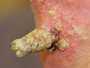
  • Cutaneous horn.
  • Actinic keratosis.
  • Keratoacanthoma.

In addition to these primary lesions, it is crucial to consider other histopathological and clinical differential diagnoses:

  • Viral wart.
  • Seborrheic keratosis.
  • Basal cell carcinoma.
  • Melanoma.
  • Blastomycosis.
  • Chondrodermatitis nodularis helicis.
Dermoscopy showing features of intraepidermal carcinoma
Dermoscopy of intraepidermal carcinoma
Macroscopic image of a prominent cutaneous horn
Macroscopic image of a cutaneous horn
Dermoscopic observation typical of an actinic keratosis
Dermoscopy of Actinic Keratosis

Dermoscopy of keratoacanthoma showing nodular features
Dermoscopy of keratoacanthoma
Dermoscopic appearance of keratoacanthoma with a keratinous center
Dermoscopy of keratoacanthoma

Correct identification of these entities through dermoscopy is crucial for establishing an appropriate management plan for squamous cell carcinoma and its clinical mimics.

Dermoscopy of a common wart showing typical features.
Dermoscopy of common wart
Dermoscopy of a viral wart.
Dermoscopy of common wart
Macroscopic image of a case of chromoblastomycosis.
Macroscopic image of chromoblastomycosis
Dermoscopic view of chondrodermatitis nodularis helicis.
Chondrodermatitis nodularis helicis dermoscopy
Dermoscopy demonstrating features of basal cell carcinoma.
Dermoscopy of basal cell carcinoma
Amelanotic melanoma with a Breslow depth of 3.2 mm, viewed with CL IV dermoscopy.
Amelanotic melanoma, Breslow 3.2 mm, CL IV dermoscopy
Dermoscopy evidencing an amelanotic melanoma.
Dermoscopy of Amelanotic Melanoma

Histological Explanation of Squamous Cell Carcinoma

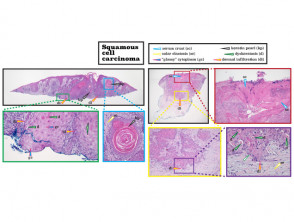
From a histological perspective, squamous cell carcinoma is characterized by the proliferation of keratinocytes atypical changes that invade the dermis.

Histopathological section showing the architecture of squamous cell carcinoma.
Histopathology of squamous cell carcinoma

Evaluating histopathology is crucial to confirm the diagnosis and determine the grade of malignancy of cutaneous malignant lesions.

Dermatly.com - El sitio de tu piel