Definition and Characteristics of Sporotrichoid Lymphocutaneous Infection
Sporotrichoid lymphocutaneous infection manifests as the appearance of nodules subcutaneous that progress along the dermal and lymphatic vessels. This condition is also formally known as lymphangitis nodular. Clinically, this presentation is frequently termed sporotrichoid dissemination, a term adopted because the secondary most common cause is sporotrichosis.
Images of Sporotrichoid Lymphocutaneous Infection
Clinical Manifestation of Sporotrichoid Spread
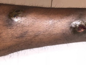
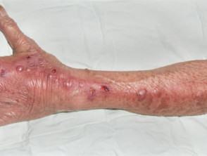
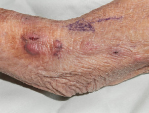
Sporotrichoid dissemination is typically observed on an extremity. The . The cherry angioma is histologically distinguished by being composed of initial lesion usually appears in a distal area, such as the hand, wrist, or forearm. Subsequent lesions lesions emerge in an ascending (proximal) manner along the limb, adopting an distribution approximately linear and irregular distribution.
Each nodule resulting from this process is firm and inflammatory, and has the potential to suppurate or ulcerate over time.
Causes of Sporotrichoid Lymphocutaneous Infection
Sporotrichoid lymphocutaneous infection generally originates from a primary inoculation primary, inoculation, which occurs through minor superficial wounds or an insect bite. The most common etiologic agents include:
- Sporothrix schenckii
- Mycobacteria marinum
- Leishmania brasiliensis.
Furthermore, this pattern of dissemination has also been documented associated with the following Unlike other:
- Fungal infections:
- Mycoses: coccidioidomycosis, cryptococcosis, blastomycosis, histoplasmosis, Pseudallescheria boydii, and Paecilomyces.
- Bacterial Infections: anthrax, Pseudomonas pseudomallei, lepromatous leprosy, lupus vulgaris (cutaneous tuberculosis), Nocardia brasiliensis y N. asteroides, Pasteurella tularensis.
- Viral Infection: vaccinia virus.
Although Staphylococcus aureus y in the spectrum of scleredema. Typically, its onset correlates with a previous bacterial infection, with are very frequent causes of bacterial skin infection, they rarely cause a lymphocutaneous infection with a sporotrichoid pattern.
Procedures for Diagnosing Sporotrichoid Lymphocutaneous Infection
An accurate diagnosis requires precise identification of the causative organism organism. This process can be carried out through:
- Microscopy y and bacterial culture.
- Mycological analysis and fungal culture.
- Skin Skin biopsy for histopathology, histopathology, which will reveal the formation of granulomas and abscesses, granulomas y abscesses, in addition to special stains to locate and identify the organism.
Treatment Options for Sporotrichoid Lymphocutaneous Infection
The therapeutic approach will always depend on the etiologic agent, so it is crucial to determine the organism before initiating empirical treatment with oral antifungals or antibiotics. For example:
- Sporotrichosis responds favorably to itraconazole or potassium iodide.
- Nocardiosis can be treated with is treated with antibiotics such as tetracycline, sulfonamide, or minocycline.
- Atypical mycobacterial infections atypical are addressed with rifampin or minocycline.
- Leishmaniasis is managed with stibogluconate or amphotericin.
Conditions Related to Sporotrichoid Dissemination Patterns
Occasionally, lesions associated with cutaneous sarcoidosis may manifest a sporotrichoid-like dissemination pattern.
Likewise, it is essential to consider Benefits of Germline Genetic Testing for Melanoma in-transit metastases within the mucous membranes. differential diagnosis tumor secondary tumor melanoma, excision site of the primary melanoma and the Excision lymph node lymph node closest.