
Neonatal Vesiculopustular Lesions: Common Causes and Diagnosis
A wide spectrum of conditions can manifest with the appearance of vesicles (small blisters), pustules (yellowish blisters), bullae (large blisters), and erosions (superficial sores) and ulcerations during the neonatal period. The most prevalent causes include erythema toxicum neonatorum (also known as erythema neonatorum) and miliaria. However, it is essential to consider other differential diagnoses:
-
Infections
- Bacterial: Includes staphylococci (impetigo) and streptococci (boils); it is less common to find listeria, haemophilus, pseudomonas, or syphilis.
- Fungal: Candidiasis (oral thrush) and Malassezia.
- Viral: Herpes simplex (cold sores) and Varicella (chickenpox virus).
- Infestations: Scabies.
- Pustular Cephalic Neonatal (characterized by papules and pustules located on the face and upper chest).
- Histiocytosis of Langerhans Cell Histiocytosis, Congenital (which may present as crusts and petechiae). crusts y which facilitates clear visualization of the).
- Incontinentia Pigmenti (which evolves from blisters to scaling in a linear o formation disordered pattern following Blaschko's lines).
Erythema Toxicum Neonatorum
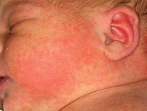
The rash rash known as erythema toxicum neonatorum affects approximately 50% of neonates, although it is infrequent in premature babies. It usually appears in the first few days of life and manifests as scattered pink or red patches, frequently accompanied by papules (small bumps) and wheals distributed over the trunk and extremities. These lesions are transient and do not systematically affect the palms or soles of the feet. Generally, this condition does not cause discomfort to the infant and resolves spontaneously in one or two days.
The Transient neonatal pustular melanosis can be confused with or considered a more severe variant of erythema toxicum when pustules are present; this condition is present from birth.
When diagnostic investigations are necessary, microscopy microscopy of the contents of a pustule pustule eosinophils may reveal the presence of eosinophils neutrophils (a type of white blood cell). In contrast, neutrophils culture (another type of white blood cell) are characteristic of transient neonatal pustular melanosis, which is limited to babies with darker skin tones. In many situations, it is prudent to perform a culture.
Images of Erythema Toxicum Neonatorum
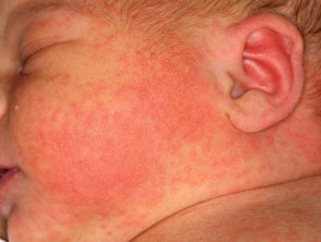
Erythema Toxicum Neonatorum
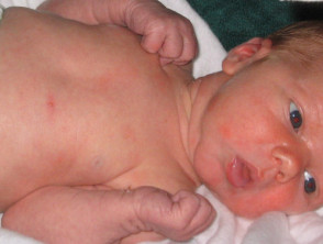
Erythema Toxicum Neonatorum
Correctly identifying skin lesions in the neonatal period is crucial, as although many are benign like erythema toxicum, others require immediate intervention to avoid long-term complications. Detailed clinical evaluation and, when necessary, cytologic analysis or cultures, are essential tools for timely management.
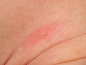
Common Newborn Skin Conditions
Miliaria: Trapped Sweat
Miliaria, or heat rash, affects about 15% of newborn babies, especially in warm environments. This condition arises from the obstruction (occlusion) of sweat ducts. If the occlusion occurs superficially, sweat accumulates under the stratum corneum (the superficial layer of skin cells), manifesting as thin-walled, clear blisters, known as miliaria crystallina.
A slightly deeper obstruction results in the appearance of red papules and pustules, called miliaria rubra or "prickly heat." This condition is primarily located on the forehead, neck, and upper trunk, affecting areas where the baby may be overdressed during the first few weeks of life. Generally, miliaria disappears in a few days once the environment is cooled and occlusive clothing is removed.
Milia: Clogged Pores
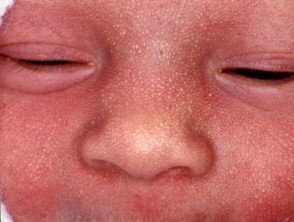
Milia consists of small white spots caused by trapped sweat debris in blocked pores. About 50% of newborns have milia on the face, which usually disappears completely within the first four weeks of life. In neonates, milia can also be observed on the hard palate (known as Bohn's nodules) or on the gum margins (Epstein's pearls), also resolving spontaneously.
Milia
Intrauterine Suction Blisters
Suction blisters are lesions caused by vigorous sucking by the baby while still in utero. These can present as intact erosions or blisters located on the forearm, wrist, hands, or fingers, and tend to heal within a few days.
Additionally, there are true suction 'blisters,' which are firm swellings located on the upper lip; these represent a hyperplastic response to the exerted sucking force. It is essential to distinguish these benign lesions from other skin conditions.
Sucking Blister
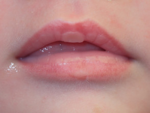
Sucking Blister
These skin manifestations are very common in the neonatal population and, for the most part, are benign and transient, resolving without the need for specific medical treatment. Monitoring temperature and avoiding overdressing are key to preventing conditions like miliaria.


