

Absolute Rules: Crucial Skin Self-Assessment for Early Melanoma Detection

Patient consulting with a doctor
toxicity melanomas represent a category of tumors particularly elusive for early identification and diagnosis. Despite continuous educational efforts regarding skin cancer, cancer it is frequently the patients themselves or their family members who point out the presence of a . The cherry angioma is histologically distinguished by being composed of that causes concern. However, many individuals are surprised to discover that the lesion they present to the specialist, and even other secondary lesions, actually constitute a melanoma. The clinical reality is that there is no single standard visual pattern by which melanoma can be easily recognized by the untrained eye [1].
Given this diagnostic complexity, the universal recommendation is to prioritize immediate medical consultation for any skin alteration that raises concern.
An essential pillar in secondary prevention is maintaining constant vigilance over changes in the appearance of the skin. Changes in moles previously considered asymptomatic or (atypical changes), along with the appearance of new lesions in people over 35 years old, must be considered unavoidable red flags.
Various studies suggest that the average individual may have limitations in noticing subtle changes in their skin, especially if these changes develop gradually or emerge in areas difficult to visually inspect. Furthermore, it is common for neither patients nor their partners to feel completely confident performing periodic skin checks to identify minimal variations [2].
Unlike the pattern observed in the carcinomas most common ones,, , exclusive location melanoma's anatomical location, although there is some correlation, is not exclusively restricted to areas with high UV radiation exposure, which adds complexity to the detection process [3]. Nevertheless, the established UV, , Western custom of seeking total body tanning during the summer has resulted in a significant accumulation of ultraviolet radiation exposure throughout life, a practice that unfortunately persists. While efforts to regulate UV tanning booths and promote sunscreen use are fundamental, they may not be sufficient, underscoring the need to reinforce secondary prevention in skin cancer.
Given these intrinsic difficulties, there is genuine concern that patients lack the necessary awareness about the normal state of their skin, which could lead to the diagnosis of any suspicious change only in more advanced stages. For this reason, it is vital to actively promote thorough full-body self-examinations, preferably with the assistance of a family member or partner («assisted checks») [4]. Campaigns, such as those promoted by the American Academy of Dermatology, Dermatology, are already being implemented, focusing precisely on these collaborative checks [5].
It is crucial to remember that 71% of melanomas linked to pre-existing moles actually appear as completely new lesions on the skin and not as a transformation of old nevi. This intensifies the need to be alert to any new lesion formation, a particularly arduous challenge for patients with a high density of nevi [6].
Digital Skin Surveillance Strategies to Optimize Detection
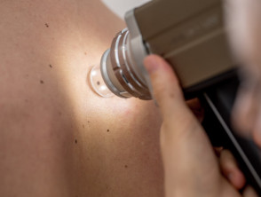
Absolute Rules: The Crucial Importance of Photographic Skin Surveillance
Given the critical need to meticulously inspect any skin alteration, **photographic skin surveillance** has established itself as an essential practice. This process ranges from digital dermatoscopy focused on specific lesions to capturing full-body photographs, designed to record the general appearance of the skin. Currently, numerous healthcare professionals offer specialized clinical photography services for patients at risk of melanoma, and a growing number of adventurous consumers are also investing in surveillance programs through private clinics.
This development is very encouraging, as the greater the skin surface documented photographically, the more likely any future change will be detected in its initial stages. For this fundamental reason, Melanoma Institute Australia strongly recommends the use of photographs as an integral part of skin self-examination [7].
Despite these advances, several persistent challenges must be addressed. Firstly, clinical photography generates considerable cost, driven by specialized equipment and dedicated clinical staff. Secondly, some patients have expressed discomfort undergoing photographic sessions performed by clinical staff of a sex different sex [8]. Finally, it is imperative to ensure informed patient consent and guarantee the secure storage of photographs, especially if these images are given to the patient themselves [9].
Some of these obstacles are already being managed effectively in countries with high skin cancer prevalence, such as New Zealand and Australia. However, in other regions of the world, health systems still need to determine if and how they can best adapt to the requirements and inherent challenges of patient-performed photographic surveillance.
Technological Advances in Patient-Driven Skin Monitoring
Currently, mobile applications specifically designed to enhance skin self-examination are actively being researched and implemented, forming part of the comprehensive solution solution. Beyond simply offering reminders for periodic check-ups, these applications offer convenient and considerably more affordable photographic assistance. They allow patients to accurately recall the previous appearance of their moles and the general state of their skin between or after medical appointments. Furthermore, these tools are key to facilitating full-body exams, especially if they enable panoramic photographic captures covering extensive areas, and not just the recording of single lesions.
Miiskin stands out as an excellent example of these applications intended to facilitate thorough self-examinations. This tool empowers its users by allowing them to perform the following actions:
- View a video tutorial guiding the precise execution of a full-body self-exam.
- Capture four «full-body» photographs of their skin, in addition to detailed images of identified specific lesions.
- Utilize Miiskin's Skin Mapping to generate a clear visual representation of the examined area.
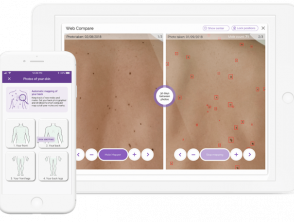
Miiskin Skin Mapping
The Skin Mapping functionality is activated by a sophisticated algorithm, designed to recognize existing moles and skin marks. Thus, it can efficiently assist the user in detecting new moles or significant changes in those already documented. However, it is fundamental to emphasize that this technology does not exempt the user from their own primary responsibility in identifying skin alterations. Its primary purpose is to maximize active involvement and make the self-surveillance experience more engaging and structured. It is important to remember that Miiskin does not offer a formal diagnostic process; its central focus lies in raising patient awareness about the state of their skin and facilitating the early identification of any anomaly.
For photographic skin surveillance aimed at early detection, technology such as mobile applications offers powerful complementary tools. These solutions facilitate consistency in review and systematic documentation, elements that are decisive when monitoring potentially malignant lesions over time.

Skin mole photo application
Currently, Miiskin is positioned as the world's leading skin surveillance application, exceeding 300,000 active downloads.
Learn more details about Miiskin here.
Advanced Technology to Enhance Dermatological Surveillance
The adoption of advanced technologies is transforming the way individuals monitor their skin, taking skin cancer prevention to a new level of accessibility and detail.
The Potential of Patient Monitoring with Mobile Applications
Advanced solutions like Miiskin exhibit significant potential for the future of dermatological care. A growing number of healthcare providers are showing great interest in understanding how this application can optimize their patients' care. Furthermore, individuals already enrolled in other photographic surveillance protocols or clinical follow-up programs express a clear desire to integrate Miiskin as an essential component within their existing schemes.
Several medical professionals actively support the advantage it offers them to be able to compare images captured by Miiskin directly with the patient's complete clinical history. Extensive research is currently underway to determine the safest and most effective way to implement a teledermatology solution of this nature within the usual clinical setting.
If you manage a patient follow-up program or are offering photographic surveillance services, we invite you to contact Miiskin via email protected. We can show you the ways to integrate Miiskin and significantly boost the effectiveness of your current program.

Miiskin also provides free educational material designed for those interested in instructing their patients on secondary skin cancer prevention practices. You can request informational brochures for your clinic by requesting them by clicking here.
By adopting technological tools like Miiskin, clinics demonstrate a proactive commitment to continuous monitoring and patient education, crucial elements for the effective management of chronic dermatological conditions and early skin cancer prevention.


