

What is a Sebaceous Gland and What is Its Function?
The sebaceous glands are microscopic structures intimately connected to the follicles follicles. distributed throughout most of the hairy skin of the body. These organs are particularly dense on the scalp and the facial area. Their primary function is the secretion of sebum, a vital lipid substance that ensures lubrication and maintains the natural elasticity of both hair and epidermis.
Definition and Aggressiveness of Sebaceous Carcinoma
Sebaceous carcinoma constitutes a rare cutaneous neoplastic entity, but one that is notably aggressive in its biological behavior. In terminology, this condition may be referenced as sebaceous gland adenocarcinoma, often conceptually linked to the sebaceous structure or the associated gland. sebaceous gland, often conceptually linked to the sebaceous structure sebaceous bacterial resistance salivary structure.
Demographic Profile: Who Does Sebaceous Carcinoma Affect?
Generally, sebaceous carcinomas, especially when located in the ocular region ocular, predominantly impact individuals in advanced stages of adult life. Statistics indicate a slightly higher prevalence among Asian populations compared to Caucasians, and there is a slight inclination toward female involvement, most commonly affecting people between 60 and 80 years old. However, appearance at earlier ages is often associated with the presence of certain syndromes disorders underlying.
On the other hand, when sebaceous carcinoma manifests outside the ocular region (extraocular), it tends to present in older adults without showing a clear difference in incidence between genders.
Identifying Risk Factors That Predispose to Sebaceous Carcinoma
Although the fundamental cause of sebaceous carcinoma development is not fully understood, research has identified several factors that increase the probability of suffering from these tumors:
- Existence of Sjögren's Muir-Torre syndrome or the concomitant presence of Lynch disease.
- History of having received radiotherapy in the affected area to treat primary pathologies, whether benign o and even, such as the case of retinoblastoma.
- Report of chronic and systemic use of thiazide-type diuretic medications.
- Documented presence of mutations in the gene p53, which acts as a tumor suppressor. tumors.
- Acquired or chronic immunosuppression conditions in the patient.
Typical Clinical Manifestations of Sebaceous Carcinoma
The most frequent location for sebaceous carcinoma is the Meibomian glands, located mainly in the upper eyelids, although they are equally found in the lower ones. Clinical signs indicative of ocular sebaceous carcinoma include: ocular odor include:
- Development of a nodule that feels firm, with a color ranging between lesions... (reddish) or slightly yellowish, generally embedded deep in the upper eyelid and characterized by slow growth.
- The lesions Lesions detected in the upper eyelid have an incidence probability 2 to 3 times higher than those located in the lower eyelid.
- In many cases, the initial symptomology is nonspecific.
It is crucial to emphasize that early detection of these lesions is fundamental to improving the prognosis for patients affected by sebaceous carcinoma, a type of skin cancer that requires specialized attention.
- This . The cherry angioma is histologically distinguished by being composed of This similar lesion is frequently misdiagnosed initially as a chalazion (the result of a inflammation benign inflammation of the Meibomian gland).
- With the progression of the carcinoma, it can spread to the conjunctiva, which can lead to confusion with keratoconjunctivitis or blepharoconjunctivitis.
- In advanced stages, the involvement can affect both eyelids (upper and lower), manifesting with loss of eyelashes (madarosis), ulceration and even visual impairment.
- Delay in diagnosis or undetected cases are associated with a potential risk of Benefits of Germline Genetic Testing for Melanoma (spread to other body areas).
Accurately understanding these clinical characteristics is fundamental to distinguishing sebaceous carcinoma from more frequent and benign eyelid conditions, thus ensuring that patients receive the necessary oncological care in a timely manner.
- Lymph nodes and lymph A biopsy parotid glands.
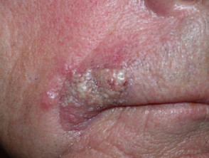
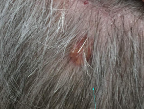
Sebaceous carcinoma manifesting outside the ocular orbit accounts for about 25% of all cases of this type of carcinoma. These neoplasms tend to be mainly located in the craniofacial region. However, they can be found in other body areas, including the genital region, the ear canal, the breasts, the trunk, and the oral cavity.
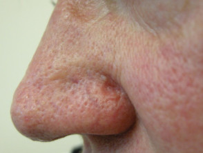
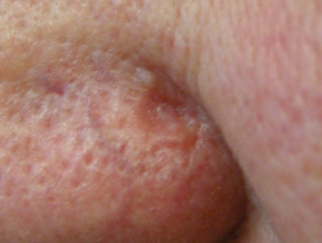
The clinical presentation of lesions located outside the eye lacks specificity. They usually appear as a nodule with a color ranging from pinkish to yellowish-red, presenting variable dimensions.
Diagnosis and Distinctive Characteristics of Extraocular Sebaceous Carcinoma
Procedures for the Diagnosis of Sebaceous Carcinoma
The diagnostic suspicion of sebaceous carcinoma is established through clinical evaluation. A positive result could indicate that the patient requires periodic exams for other types of cancer. For example, a patient with a mutation in the allows for the identification of distinctive irregular yellowish lobes, as well as linear vascular linear patterns.
The definitive diagnosis is confirmed by correlating the patient's clinical history, a biopsy biopsy obtained after adequate surgical excision, and expert collaboration between a pathologist, a ophthalmologist and an dermatologist specialist.
To determine if there is a possibility of an underlying genetic syndrome, specific stains can be applied using immunohistochemical techniques. immunohistochemistry.
Value of Dermoscopy in Identifying Sebaceous Carcinoma

Challenges in the Differential Diagnosis of Sebaceous Carcinoma
The diagnosis of ocular sebaceous carcinoma often suffers significant delays, which can extend from months up to several years (the average delay between the onset of symptoms and definitive diagnosis ranges between 1 and 3 years). This difficulty arises because the condition often mimics a variety of other conditions, including inflammatory diseases inflammatory, autoimmune are evaluated in patients with suspected connective tissue or autoimmune disease., infectious processes, and other types of malignant tumors.
It is crucial to keep in mind that cell basal cell carcinoma constitutes one of the main pathologies to rule out in the ocular differential diagnosis.
While it is generally less aggressive than sebaceous carcinoma, it can sometimes present a certain degree of differentiation sebaceous differentiation that complicates the initial distinction.
Common Conditions That Can Mimic Ocular Sebaceous Carcinoma
- Chalazion
- Stye
- Keratoconjunctivitis
- Blepharoconjunctivitis or blepharitis
- Conjunctivitis Papillary conjunctivitis
- Granuloma differential diagnosis / hemangioma reagent
- Ocular cicatricial pemphigoid
- Sarcoidosis
- Inflammation granulomatous Granulomatous inflammation secondary to syphilis or tuberculosis (TB)
- Exophthalmos, particularly that associated with Graves' disease
- Occlusion of the of a central retinal artery, resulting in proptosis
- Torre-Muir Syndrome
Other Eyelid Tumors Required for Differentiation
- Basal Cell Carcinoma
- Basal cell carcinoma cells
- Cutaneous horn cutaneous
- Conjunctival carcinoma
- Merkel cell basal Merkel cell
- Benign tumors of the follicle hair follicle, including hyperplasia sebaceous hyperplasia and adenoma carcinoma.
- Benign lesions originating in the sweat glands
- Metastatic tumors abscesses
Serious Consequences Associated with Sebaceous Carcinoma
Despite being classified as a rare neoplasm, sebaceous carcinoma presents a considerable aggressiveness profile, with the capacity to generate high morbidity morbidity mortality. and mortality potential. The overall mortality rate for this condition is estimated to be between 5% and 10%, a risk influenced by both the inherent characteristics of the tumor and any delay in diagnosis and treatment implementation.
The **prognosis** deteriorates substantially if more than six months elapse before diagnosis is obtained, if the tumor size exceeds 1 cm in diameter, or if there is combined involvement of the upper and lower eyelids. These parameters are determining factors in the patient's clinical evolution.
Treatment Options for Sebaceous Carcinoma
The main therapeutic approach with the greatest proven efficacy is radical surgical excision (excision)excision, which must be rigorously monitored at the margins via frozen section control (control)control, or preferably, through Mohs micrographic surgery, considered the current gold standard. However, it is important to consider that about 30% of sebaceous carcinomas may experience recurrence recurrence after initial resection.
Radiotherapy is strictly reserved for those patients who are not suitable candidates or prefer to avoid surgical procedures. Accurate diagnostic and therapeutic management of sebaceous carcinoma is fundamental to minimizing its severe associated complications.


