
Sclerosing Lipogranuloma: Clinical Manifestations and Histopathological Insights
Sclerosing lipogranuloma frequently presents as a palpable induration, either painless or slightly tender, typically located in the penis, scrotum, spermatic cord, or perineum. This pathological entity usually arises as a consequence of the injection or topical application of oily agents, such as paraffin, silicone, oil, or wax, introduced for aesthetic or therapeutic purposes.
Histopathological Characteristics of Sclerosing Lipogranuloma
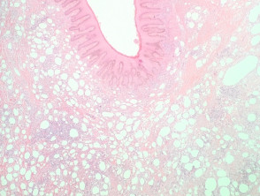
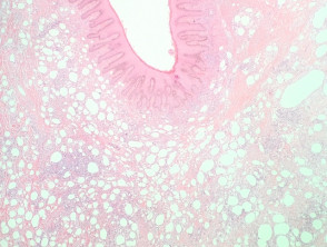
Histopathological analysis of sclerosing lipogranuloma reveals distinctive findings, including fat necrosis, histiocyte accumulation, and the presence of multinucleated giant cells. These elements coexist with marked fibrosis and signs of tissue hyalinization. It is common to observe polymorphous vacuoles containing the foreign material and the subsequent formation of pseudocysts, areas with a higher concentration of the injected exogenous substance (see Figures 1 and 2).
Detailed Analysis of Sclerosing Lipogranuloma Pathology
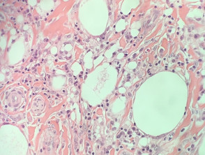
Generally, routine diagnosis does not require special stains for confirmation. Although Oil Red O staining on frozen tissue is positive for lipids, its practical application in the clinical setting is infrequent.
Key Strategies for the Differential Diagnosis of Lipogranuloma
Establishing an accurate diagnosis of lipogranuloma requires careful differentiation from other pathologies that present similar morphological characteristics. The main differential diagnoses to consider include:
- Liposarcoma: This represents the most significant differential diagnosis that must be ruled out. Histologically, liposarcoma is distinguished by the presence of adipocytes of heterogeneous size, the identification of lipoblasts, and, crucially, the notable absence of multinucleated giant cells.
- Lymphangioma: The distinction becomes more complex when lymphangiomas have been subjected to prior treatment with sclerotherapy. Unlike the pseudocysts characteristic of sclerosing lipogranuloma, lymphangiomas are defined by the presence of endothelium-lined lymphatic spaces.
To ensure appropriate clinical management, it is essential to correlate clinical findings with histopathological results, which confirms the diagnosis of sclerosing lipogranuloma as a tissue reaction secondary to the introduction of foreign substances into the body.


