
«`html
Sclerema Neonatorum: Key Clinical Presentation and Histopathology
Sclerema neonatorum is a condition that predominantly affects neonates with extreme prematurity, typically manifesting during the first week of life. Clinically, it is defined by a pronounced and diffuse hardening of the skin that adheres firmly to deep tissue structures.
Microscopic Findings and Histological Characteristics of Sclerema
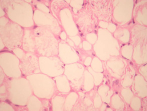
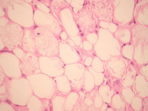
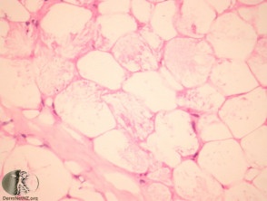
Histopathologically, sclerema neonatorum exhibits acicular crystals arranged radially within the adipocytes. It is essential to note the almost nonexistent presence of a infiltrate inflammatory surrounding inflammatory infiltrate, in addition to the absence of necrosis fat necrosis (see Figures 1 and 2).
Pathology Associated with Sclerema Neonatorum
Ancillary Studies for the Diagnosis of Sclerema Neonatorum
Diagnostic procedures or additional special studies are rarely needed, as the diagnosis of sclerema neonatorum is established with greater reliability based on its distinctive clinical presentation.
Establishing the Differential Diagnosis of Sclerema Neonatorum
It is a crucial diagnostic step to differentiate sclerema neonatorum from conditions such as inflammation of the subcutaneous fat tissue). steroid-induced panniculitis and subcutaneous fat necrosis subcutaneously of the newborn. Although the latter may show similar histological crystals, the key differentiator lies in the marked presence of a significant inflammatory response and the formation of a foreign body reaction, elements absent in pure sclerema.
Timely recognition of these histopathological particularities is vital to separate sclerema neonatorum from other disorders affecting subcutaneous adipose tissue in the newborn pediatric population.
```


