
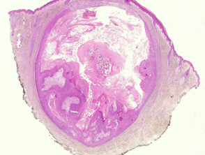
Fundamental Rules for the Accurate Diagnosis of Proliferating Epidermoid Cyst
The definition of proliferating epidermoid cyst has historically been a topic of debate in the medical literature. To establish an unequivocal diagnosis, this cyst variant must exhibit, at least partially, the characteristic cystic structure along with a proliferation evident epithelium underlying.
Detailed Histological Analysis of Proliferating Epidermoid Cyst
Upon examining the histopathological samples of this type of cyst, a lesion localized in the dermis exhibits an active and constantly growing epithelial component. A key sign in these proliferative areas proliferative is the presence of characteristic bland squamous epithelium, where squamous eddies are notable. These eddies are, in fact, whorls of squamous epithelium in the process of maturation, showing morphology identical to that observed in irritated seborrheic keratosis or in lesions of seborrheic keratosis irritated or in lesions of queratosis folicular inverted keratosis.
Histopathological Correlation: Images of Proliferating Epidermoid Cyst
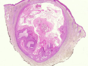
Figure 1
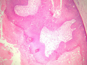
Figure 2
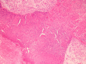
Figure 4
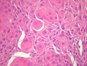
Figure 4
Complementary Evaluations for Proliferating Epidermoid Cyst
In clinical practice, it is rarely necessary to resort to advanced complementary studies, such as molecular biopsies or special stains, to confirm the diagnosis of proliferating epidermoid cyst. Diagnostic confirmation is fundamentally based on the rigorous interpretation of the standard histopathological evaluation.
Establishing the Differential Diagnosis in Pathology of Proliferating Cysts
It is essential to differentiate this clinical entity from other cystic lesions of epidermal lineage. A key differential diagnosis involves epidermal cysts associated with HPV, which usually show a hyperplastic lining epithelium accompanied by nuclear and cytoplasmic alterations with evident viropathic features.
Another diagnosis that warrants exhaustive consideration is cystic squamous cell carcinoma. This malignancy must be ruled out with special attention whenever marked cellular atypia and evidence of infiltration into the dermis adjacent. This differentiation is significantly complicated in cases where the proliferative portion is extensive or there has been a partial cystic rupture.


