
Histologic Diagnosis of Pilomatricoma
Pilomatricoma is a cutaneous neoplasm that originates, according to current evidence, from the hair matrix cells.
Key Histologic Features of Pilomatricoma
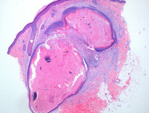
At low magnification, the histopathological level characteristic histopathologic pattern of pilomatricoma is evident as a well-circumscribed nodulocystic tumor circumscribed (Figure 2). Although it is located mainly in the deep dermis dermis, it is not unusual for it to extend into the subcutaneous tissue. subcutaneously.
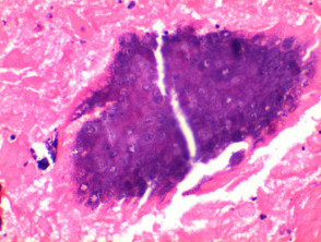
Structurally, this tumor is composed of a proliferation of basaloid cells basaloid that emulate hair matrix cells. These cells undergo maturation into eosinophilic structures, eosinophilic, which lack nuclei nuclei. and are known as shadow cells (Figures 3 and 4). This area of shadow cells reflects differentiation differentiation toward the cortex of the hair follicle. It is common to observe foci of calcificación calcification within these shadow cell regions (Figure 5). Furthermore, it is common to find a infiltrate histiocytic infiltrate that includes multinucleated cells multinucleated cells. at points of tissue rupture (Figure 6).
Illustrations of Pilomatricoma Pathology
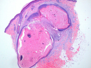
Figure 1
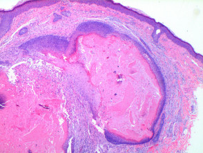
Figure 2
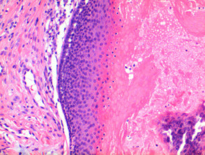
figure 3
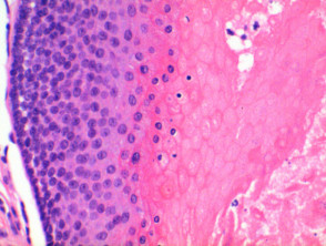
Figure 5
Special Stains and Syndromes Associated with Sebaceoma

Figure 6
Use of Special Stains in the Diagnosis of Pilomatricoma
Generally, the application of special stains is not required to confirm the histologic diagnosis of pilomatricoma. However, Von Kossa special stain can be useful to highlight and confirm the presence of calcium deposits in the tissue sample.
Understanding the histology is fundamental to differentiating pilomatricoma from other adnexal skin tumors, ensuring adequate clinical management.


