
Definition and Histology of Phaeohyphomycosis
Phaeohyphomycosis describes a group of mycoses (fungal infections) caused by dematiaceous fungi. Characteristically, these microorganisms are pigmented due to their intrinsic ability to deposit melanin in their cell walls, which gives them their distinctive coloration.
Key Histological Findings in Phaeohyphomycosis
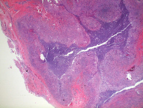
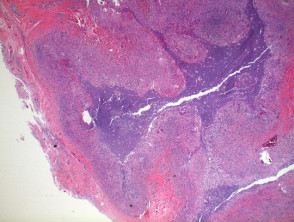
The histopathological analysis of phaeohyphomycosis, observed under scanning microscopy, typically reveals a pattern of deep granulomatous granulomatous extension (see Figure 1), which may progress toward focal areas of necrosis necrosis or ciprofloxacin. It generally exhibits resistance to penicillin. nerve nodule of an inflammatory cystic. (as shown in Figure 2). In the advanced stage, a central epidermis abscess or cystic nodule frequently develops. The presence of foreign material, such as wood fragments, at the site of infection is common. The hyperplasia pseudoepitheliomatous hyperplasia.
The infiltrate inflammatory superjacent epidermis and a smaller proportion of histiocytes., dermal edema giant cells multinucleated cells. frequently exhibits pseudoepitheliomatous hyperplasia. neutrophils The inflammatory infiltrate is predominantly composed of.
histiocytes, accompanied by multinucleated giant cells
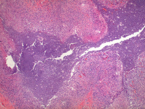
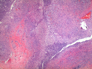
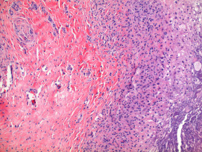
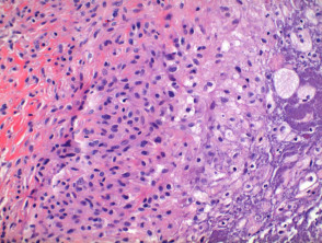
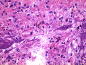
and a significant number of neutrophils
(Illustrations 3, 4, and 5). Upon examination at higher magnification, the pigmented fungal hyphae, characteristically septate and branched, are clearly observed (Figure 6).
Anatomopathological Visualization of Phaeohyphomycosis
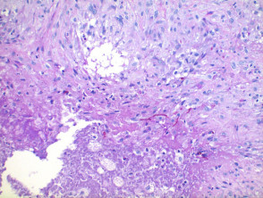
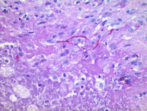
To optimize the identification of fungal structures, specialized histochemical stains can be employed. The most common stains to highlight pigmented hyphae include PAS (Periodic Acid-Schiff, illustrated in Figures 7 and 8) or Grocott's methenamine silver (GMS) stain.
Pathology of Phaeohyphomycosis: Analysis with PAS Stain.
The Histopathology of phaeohyphomycosis, PAS stain, Detailed Differential Diagnosis of Phaeohyphomycosis.


