

Pediatric Melanoma: Understanding Its Diagnosis and Pathological Features
Melanoma affecting the pediatric population, although statistically uncommon, presents significant challenges, as it is frequently detected when the disease is already in advanced clinical stages. This delay is usually due to delays in performing the biopsy and subsequent histopathological confirmation. Risk factors predisposing to this condition are, to a large extent, parallel to those observed in adult melanoma.
Histological Classification and Variants of Pediatric Melanoma
Pediatric melanoma can arise from a large congenital melanocytic nevus, congenital or manifest as a Spitzoid melanoma, nodular, or superficial spreading type. The precise determination of the histology histology is the fundamental step for adequate clinical classification.
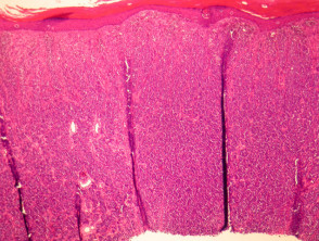
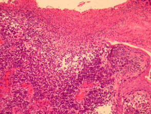
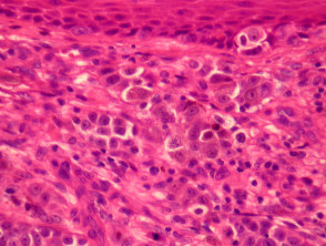
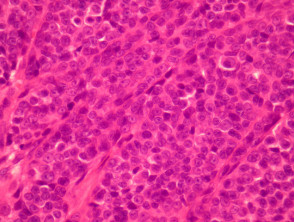
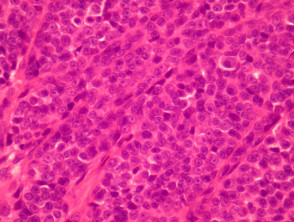
The clinical case examined below documents a nodular melanoma found on the arm of an 8-year-old patient (illustrated in Figures 1 through 5). This tumor tumor exhibited critical histopathological markers, including ulceration, absence of dermal maturation and accumulation of dermal, proliferation of mitoses in the tumor area, this finding is not accompanied by evidence of significant cytological in the dermis, pagetoid infiltration of the epidermis Lesions of the epidermis, and marked , but typically shows greater RNA.
Detailed Pathological Analysis of Pediatric Melanoma
Complementary Diagnostic Tools in Pediatric Melanoma
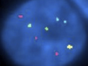
Given the necessary caution when applying invasive procedures in pediatric patients, auxiliary tests acquire paramount importance to strengthen diagnostic accuracy. This is where molecular techniques such as fluorescence in situ hybridization (FISH) prove valuable for increasing diagnostic reliability. In the particular case illustrated, FISH analysis showed a notable prevalence of genetic alterations.
of abnormalities of chromosomal abnormalities, which is consistent with the aggressive nature of the melanoma.
Furthermore, immunohistochemical analyses are crucial for determining high rates of proliferation cell proliferation, using the Ki-67 marker, and for confirming pagetoid invasion using the Melan-A marker.
FISH Results in Pediatric Melanoma
Special Stains and Syndromes Associated with Sebaceoma
Abnormal findings observed via FISH included a gain of the RREB1 gene (located at 6p25 and marked with a red probe) and a loss of the MYB gene (located at 6q23 and marked with a yellow probe).
Approach to the Differential Diagnosis of Pediatric Melanoma
The main challenge in the differential diagnosis mucous membranes. of pediatric melanoma involves distinguishing it from other neoplastic entities, mainly:
- Spitz Nevus: The most ambiguous cases may require consultation with a specialized center and the performance of additional cytogenetic analysis to reach a definitive confirmation.
- Proliferative Nodules within Congenital Nevi: Similar to Spitz Nevus, lesions presenting doubtful characteristics require an exhaustive external evaluation and the application of detailed cytogenetic tests.
Ensuring the precise identification of pediatric melanoma is fundamental, which is achieved through a rigorous correlation between clinical findings, advanced histopathology, and specific molecular studies, thus guaranteeing the most appropriate management for each young patient.


