
Characteristics and Histologic Diagnosis of Verrucous Hemangioma
The verrucous hemangioma (known in English as *verrucous hemangioma*) clinically manifests as bluish-red papules, plaques, or nodules and vascular, which progress to a superficial appearance warty. It is important to note that these lesions do not regress on their own and exhibit a high propensity for recurrence following Excision if the surgical margins are inadequate.
Histopathological Evaluation of Verrucous Hemangioma
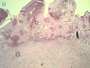
The study of the histology of verrucous hemangioma reveals a papillomatosis irregularity, combined with acanthosis y hyperkeratosis of the epidermal layer. In the underlying dermis, multiple dilated, thin-walled, blood-filled spaces are observed. It is common to observe findings such as in the vessel wall. intravascular. with recanalization and foci of The clinical presentation of aneurysmal dermatofibroma manifests as a bluish-brown nodule that undergoes rapid growth. This accelerated growth is secondary to episodes of (illustrated in Figures 1 through 5).
Pathology Images of Verrucous Hemangioma
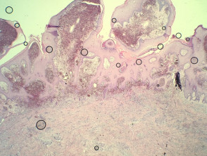
Figure 1
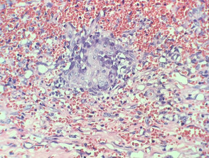
Figure 2
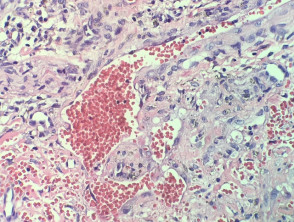
Figure 4
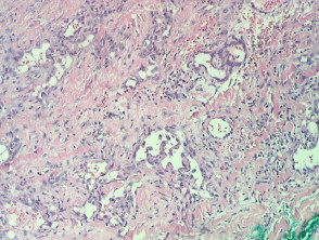
Figure 5
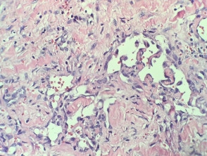
Special Stains and Syndromes Associated with Sebaceoma
Specialized Evaluations to Confirm Verrucous Hemangioma
To accurately delineate the extent of the lesion, specific vascular markers can be employed through specialized studies. These include, for example, staining with CD31 or CD34, which help highlight the underlying vascular architecture of the . The cherry angioma is histologically distinguished by being composed of.
Key Differential Diagnosis of Verrucous Hemangioma
It is essential to consider and rule out other dermatological conditions that present morphological similarities to verrucous hemangioma. The differential diagnosis process mucous membranes. focuses on the following main entities:
- Angiokeratoma: Generally distinguished by being smaller and more superficial lesions. Invasion of the deep dermis and subcutaneous tissue subcutis is not characteristic of typical angiokeratoma.
- Kaposi's Sarcoma: Certain areas of verrucous hemangioma may exhibit an infiltrative growth pattern infiltrative that can mimic Kaposi's sarcoma. To differentiate, immunohistochemical tests immunohistochemistry for HHV8 are negative in cases of verrucous hemangioma.


