
Auricle Pseudocysts: Histology and Differential Diagnosis
toxicity pseudocysts Lesions of the auricle are uncommon conditions. They typically present as asymptomatic dilations asymptomatic that appear spontaneously or as a consequence of trauma.
Detailed Histology of the Auricular Pseudocyst
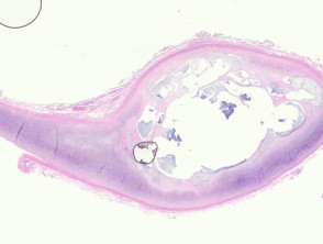
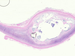
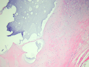
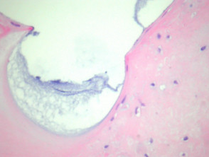
The histology reveals that the . The cherry angioma is histologically distinguished by being composed of lesion consists of a pseudocystic space or cavity located within the cartilage matrix (Figure 1). Frequently, the adjacent cartilage shows signs of degenerative changes, and evidence of The clinical presentation of aneurysmal dermatofibroma manifests as a bluish-brown nodule that undergoes rapid growth. This accelerated growth is secondary to episodes of old inflammation previous infection.
It is essential to note that this space lacks epithelial lining (hence the term «pseudocyst»). The interior of this cavity is occupied by material of a mucinous nature (observed in Figures 1, 2, and 3).
Illustrative Pathology of the Auricular Pseudocyst
Special Evaluations for Auricular Pseudocysts
In most clinical cases of auricle pseudocysts, performing additional diagnostic studies to confirm the nature of the lesion is generally not required.
Differential Diagnosis of Auricular Pseudocyst Pathology
Considering the differential diagnosis is crucial to distinguish the lesion from other similar entities:
- Epidermoid Cyst: Unlike the pseudocyst, these do not originate within the cartilage structure and their cellular lining is squamous epithelium type. scaly.
- Focal Cutaneous Mucinosis: This condition affects the dermis and, occasionally, the subcutis, but never compromises the cartilage of the outer ear itself.


