
Lymphomatoid Papulosis: Classification and Histological Characteristics
The lymphomatoid papulosis is classified as one of the two lymphomas skin's blood vessels melanomas CD30-positive, according to the diagnostic criteria of the World Health Organization (WHO/EORTC).
Detailed Histology of Lymphomatoid Papulosis
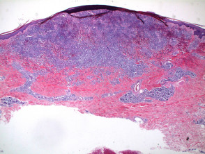
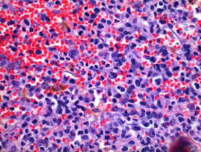
The histopathological analysis of lymphomatoid papulosis reveals a wedge-shaped inflammatory infiltrate that extends deep into the dermis or into the subcutaneously superficial subcutaneous tissue (as illustrated in Figures 1 and 2). It is important to note the presence of telangiectatic prominent extravasation of erythrocytes, visible in Figures 3 and 4, in the case presented.
The population lymphocytic consists predominantly of lymphocytes large cells positive for CD30 (Figure 5). This cellular mixture forms a spectrum that is classified into several types:
- Type A: It is characterized by large CD30-positive lymphocytes, which appear scattered or clustered (Figure 6). This pattern also includes a background with a predominance of eosinophils y neutrophils.
- Type B: It shows a population composed of small lymphocytes exhibiting is observed. This subtype may present morphological similarities to mycosis fungoides.
- Type C: Sheets of large anaplastic,, anaplastic cells are observed, with very few mixed inflammatory cells. This pattern evokes the morphology of a lymphoma large cell anaplastic lymphoma.
- Type D: It is characterized by marked epidermotropism, being histologically indistinguishable from CD8+ epidermotropic cutaneous T-cell lymphoma.
Images of Lymphomatoid Papulosis Pathology

Figure 1
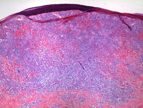
Figure 2
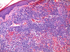
Figure 4
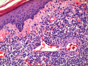
Figure 5
Special Stains and Syndromes Associated with Sebaceoma
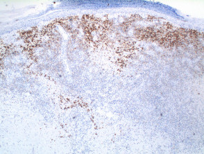
Special Stains in Lymphomatoid Papulosis
Evaluation requires a panel of markers via immunoperoxidase. The crucial finding is CD30 positivity in subtypes A and C. Typically, the tumor cells tumor are CD4+, although CD8+ and CD56+ variants have been documented. Expression of TIA-1, perforin, and granzyme B is common, while ALK-1 is negative.
Additionally, Fascin expression may indicate an increased risk of developing secondary malignant neoplasms.
Differential Diagnosis of Lymphomatoid Papulosis
The main differential diagnosis includes Primary Cutaneous Anaplastic Large Cell Lymphoma (PCALCL). The latter presents clinically in a distinct manner, resembling a large, solitary tumor, often ulcerated. By strict definition, more than 75% of tumor cells must show CD30 positivity in PCALCL. Furthermore, it is characteristic to observe a cohesive population of large anaplastic cells, frequently with a nodular prominent pattern.
Accurate diagnosis requires correlating immunohistochemical findings with clinical presentation and differential histological morphology to rule out these related malignant entities.


