Introduction to Erosive Papillomatosis of the Nipple
This condition, also known as florid nipple, papillary papillomatosis, or papillary adenoma, manifests as erythema or nodules on the nipple. Visually, it can be confused with mammary Paget's disease, which requires correct diagnostic differentiation.
Detailed Histology of Erosive Papillomatosis of the Nipple and Papillary Adenoma
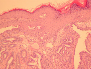
Histological analysis of erosive papillomatosis of the nipple reveals a papillary epithelial proliferation that connects with the overlying epidermis (Figure 1). The lesion is structurally composed of fibrovascular cores that are lined by cells with a bland appearance (Figure 2).
Characteristically, these cells usually present at least two layers thick, composed of a myoepithelial layer and an epithelial layer. These layers frequently show apocrine differentiation (Figure 3).
Pathological Findings of Erosive Papillomatosis of the Nipple

Figure 1
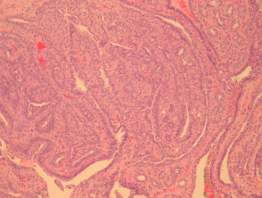
Figure 2
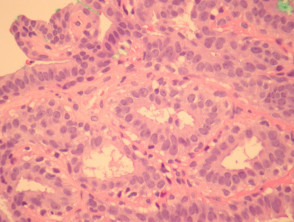
Figure 4
Clinical Investigation and Special Studies
Generally, specific complementary studies are not required to confirm the diagnosis of erosive papillomatosis of the nipple once the biopsy for histopathological study has been performed.
Crucial Differential Diagnosis
It is essential to differentiate erosive papillomatosis of the nipple from other malignant entities, especially carcinoma. Breast carcinoma infiltrating the nipple will exhibit much more marked nuclear atypia. Furthermore, malignant neoplasms typically lack the organized papillary growth pattern and, crucially, do not present the double lining composed of myoepithelial and epithelial cells that characterizes benign lesions.