
Histology and Pathology of Elastosis Perforans Serpiginosa
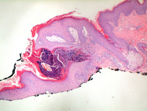
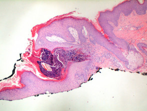
Low-power histological observation of elastosis perforans serpiginosa (EPS) reveals a column of keratotic material that forms a focal invagination through a hyperplastic epidermis (Figure 1). Closer examination allows identification of the material in the process of elimination transepidermally (Figure 2).
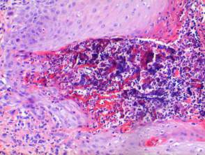
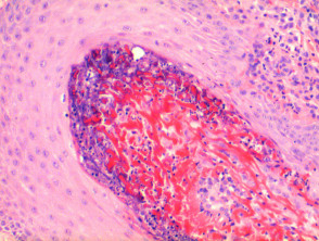
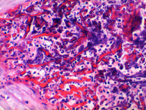
Brightly stained fibers of eosinophil are seen within the extruded material, mixed with keratinous debris and a dense inflammatory infiltrate of cells (Figures 3 and 4). These findings are crucial for the definitive diagnosis of this dermatosis.
Detailed Pathology of Elastosis Perforans Serpiginosa
Use of Special Stains in Elastosis Perforans Serpiginosa
To confirm the nature of the material involved, special stains are used. The Elastic-Van Gieson (EVG) protocol notably highlights the elastic fibers elastic fibers, which appear black as they cross the focus of transepidermal elimination (Figures 5 and 6).
Evidence of Elastic Fibers with Elastic-van Gieson Stain
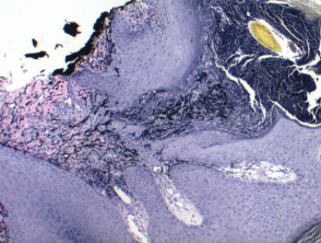
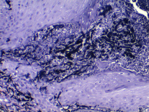
Rigorous histopathological analysis, supplemented with stains such as EVG, is fundamental for differentiating elastosis perforans serpiginosa from other skin conditions that present alterations in elastic connective tissue.
«`html
Differential Diagnosis of Elastosis Perforans Serpiginosa
The differential diagnosis of elastosis perforans serpiginosa (EPS) requires careful distinction from other skin conditions that present transepidermal material.
Traumatic Collagen Perforation
This presentation is characterized by a traumatic erosion of the epidermis, where altered collagen fibers on the dermal surface may appear to be passing through (perforating) the residual epithelium. It is important to note that this secondary phenomenon is not uncommon in lesions associated with prurigo nodularis. The presence of extensive areas of epidermal erosion, along with adjacent epidermal hyperplasia and vertically oriented dermal fibrosis, are key indicators of an underlying chronic prurigo, suggesting the etiology of the condition. A relevant histopathological finding is that the extruded fibers in this context correspond to collagen and will therefore test negative with EvG (Elastin-Van Gieson) stain.
Kyrle's Disease
In Kyrle's Disease, the material observed "perforating" the skin consists strictly of inflammatory remnants from the dermis, and not elastic tissue or collagen. It is crucial to differentiate it from reactive perforating collagenosis due to possible diagnostic overlaps.
Reactive Perforating Collagenosis
Unlike EPS, in reactive perforating collagenosis, the fibers extruded through the epidermis are collagenous in nature and will show negativity to EVG staining.
The correct identification of the nature of the transepidermal material, along with the surrounding histopathological features, is essential for establishing an accurate diagnosis and differentiating EPS from these related conditions.
```


