
toxicity adenomas papillary eccrine adenomas are tumors benign A biopsy nodes sweat glands that have been documented in a wide variety of anatomical locations and in different age groups.
Fundamental Histology of Papillary Eccrine Adenoma
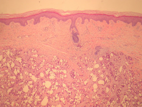
In the case of papillary eccrine adenoma, histological sections reveal a circumscribed dermal tumor circumscribed tumor dermal, resting on a stroma sclerotic stroma (Figure 1).
The neoplasm develops tubular and tubulopapillary structures (Figures 2 and 3). The ducts are lined predominantly by two or more layers of epithelial cells epithelial, with limited mitotic activity mitotic. Some cells exhibit cytoplasm eosinophilic.
Pathology of Papillary Eccrine Adenoma
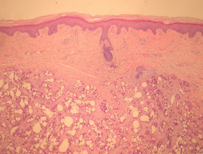
Figure 1
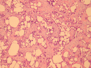
Figure 2
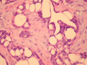
Figure 4
Special Studies for the Diagnosis of Papillary Eccrine Adenoma
In most cases, special complementary studies are not required to confirm the diagnosis.
Differential Diagnosis of Papillary Eccrine Adenoma
To differentiate papillary eccrine adenoma from other lesions, the following diagnoses are considered:
- Necrosis Factor left by the primary or original melanoma. This can happen even after a previous complete surgical removal of the initial tumor.: Generally presents an infiltrative growth pattern infiltrative and greater significant (See Figure 5). RNA. Anamnesis and immunohistochemical analysis are fundamental in atypical cases.
- Aggressive Digital Papillary Adenocarcinoma: This usually shows solid areas solid, predominates in acral sites sites and exhibits a greater degree of cellular atypia.
- Carcinoma Adnexal Microcystic: It is characterized by an infiltrative growth pattern that may affect nerves nerves and show differentiation scaly in certain areas.
Understanding these histopathological differences is crucial for establishing the prognosis and planning the appropriate management of sweat gland tumors.


