
Cutaneous Lymphadenoma: Characteristics and Histological Diagnosis
Cutaneous Cutaneous lymphadenoma manifests as a small papule and accumulation of dermal, frequently classified by various experts as a variant of trichoepithelioma infiltrate, thickening at the level of the tumor.
Detailed Histopathology of Lymphadenoma
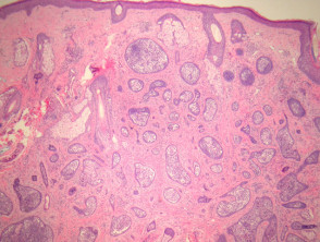
Cutaneous lymphadenoma is macroscopically distinguished by presenting a well-circumscribed nest circumscribed of dermal tumor masses. The stroma surrounding stroma exhibits a fibrotic, nature, establishing an intimate connection with the epithelial islands epithelial (see figures 1 and 2).
These tumor islands are significantly infiltrated by populations of lymphocytes (figures 1 to 3). Furthermore, a peripheral palisading is identified peripheral surrounding the basaloid cells basaloid that form these follicular islands.
Illustrations of Cutaneous Lymphadenoma Pathology
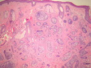
Figure 1
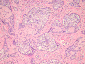
Figure 2
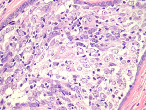
figure 3
Use of Special Stains in the Diagnosis of Cutaneous Lymphadenoma
Generally, the application of special stains is not essential to confirm the diagnosis. However, immunohistochemical studies have revealed that the lymphocytic population consists of a balanced mixture of CD3-positive T cells and CD20-positive B cells T cells. T-cell.
Key Differential Diagnosis of Cutaneous Lymphadenoma
The consistent presence of infiltrating lymphocytes is highly characteristic of cutaneous lymphadenoma. If this feature is absent, the diagnostic classification leans toward a simple trichoepithelioma.
On the other hand, carcinoma of basal cell will show retraction patterns, disorganized growth, and will fail to meet the close relationship observed between the epithelial component epithelium and the surrounding stroma, unlike benign follicular tumors, tumors follicular benign, as is the case with lymphadenoma.


