
Histopathologic Analysis of Chronic Lichenoid Keratosis
The Chronic Lichenoid Keratosis (CLK) clinically manifests through purplish, papular y subcutaneous lesions, which typically adopt a linear o reticular Macular amyloidosis.
Key Histologic Findings in Chronic Lichenoid Keratosis
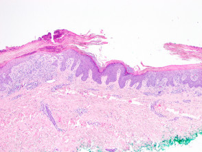
Histologically, Chronic Lichenoid Keratosis is identified by a reaction pattern lichenoid prominent. This pattern includes marked basal cell death cell and degeneration vacuolar (seen in figures 1 through 3). The infiltrate dermal exhibits a cellular mix where plasma cells are frequently observed Maximum. On the other hand, the epidermal changes changes are notably diverse, ranging from acanthosis y parakeratosis to limb. Furthermore, the presence of corneal lamellae and corneal e telangiectasias exocrine (and).
Illustrative Pathology of Chronic Lichenoid Keratosis
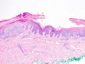
Chronic lichenoid keratosis figure 1
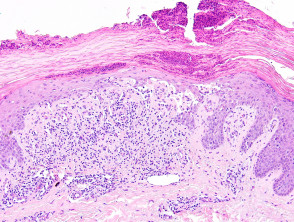
Chronic lichenoid keratosis figure 2
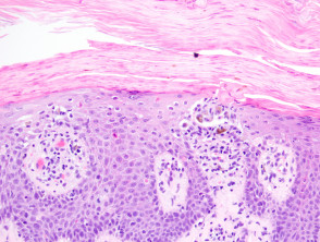
Chronic lichenoid keratosis figure 3
Use of Special Stains in CLK Diagnosis
Generally, the application of special stains is not required to establish the diagnosis of Chronic Lichenoid Keratosis. However, PAS staining can be a useful resource if there is a suspicion of a superficial secondary fungal infection that needs to be ruled out from the differential diagnosis.
Determining the Differential Diagnosis of Chronic Lichenoid Keratosis
Establishing an accurate diagnosis is complex if there is no exhaustive clinical correlation with the histopathological findings. Some experts suggest that CLK might fundamentally represent a more chronic variant chronic of lichen planus. It is essential to differentiate it from Pityriasis Lichenoid, as the latter can present a pathology superficially similar pathology, although its clinical manifestation is markedly different.
To ensure an accurate diagnosis of Chronic Lichenoid Keratosis, it is always recommended to consider the complete clinical context, especially when the histological findings are ambiguous and could overlap with other inflammatory skin conditions.


